 Movie
Movie Controller

[English] 日本語
 Yorodumi
- PDB-1pdl: Fitting of gp5 in the cryoEM reconstruction of the bacteriophage ... -
+ Open data

- Basic information
| Entry | Database: PDB / ID: 1pdl | ||||||
|---|---|---|---|---|---|---|---|
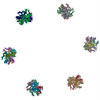
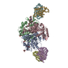
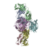
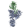
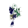
| Title | Fitting of gp5 in the cryoEM reconstruction of the bacteriophage T4 baseplate | ||||||
 Components Components | Tail-associated lysozyme | ||||||
 Keywords Keywords |  HYDROLASE HYDROLASE | ||||||
| Function / homology |  Function and homology information Function and homology informationsymbiont entry into host cell via disruption of host cell wall peptidoglycan / virus tail, baseplate / viral tail assembly / symbiont entry into host cell via disruption of host cell envelope / virus tail / symbiont entry into host / peptidoglycan catabolic process / cell wall macromolecule catabolic process / lysozyme / lysozyme activity ...symbiont entry into host cell via disruption of host cell wall peptidoglycan / virus tail, baseplate / viral tail assembly / symbiont entry into host cell via disruption of host cell envelope / virus tail / symbiont entry into host / peptidoglycan catabolic process / cell wall macromolecule catabolic process / lysozyme / lysozyme activity / killing of cells of another organism / defense response to bacterium / symbiont entry into host cell / identical protein bindingSimilarity search - Function | ||||||
| Biological species |  Enterobacteria phage T4 (virus) Enterobacteria phage T4 (virus) | ||||||
| Method | ELECTRON MICROSCOPY / single particle reconstruction / cryo EM / Resolution: 12 Å | ||||||
 Authors Authors | Kostyuchenko, V.A. / Leiman, P.G. / Chipman, P.R. / Kanamaru, S. / van Raaij, M.J. / Arisaka, F. / Mesyanzhinov, V.V. / Rossmann, M.G. | ||||||
 Citation Citation | Journal: Nat Struct Biol / Year: 2003 Title: Three-dimensional structure of bacteriophage T4 baseplate. Authors: Victor A Kostyuchenko / Petr G Leiman / Paul R Chipman / Shuji Kanamaru / Mark J van Raaij / Fumio Arisaka / Vadim V Mesyanzhinov / Michael G Rossmann /  Abstract: The baseplate of bacteriophage T4 is a multiprotein molecular machine that controls host cell recognition, attachment, tail sheath contraction and viral DNA ejection. We report here the three- ...The baseplate of bacteriophage T4 is a multiprotein molecular machine that controls host cell recognition, attachment, tail sheath contraction and viral DNA ejection. We report here the three-dimensional structure of the baseplate-tail tube complex determined to a resolution of 12 A by cryoelectron microscopy. The baseplate has a six-fold symmetric, dome-like structure approximately 520 A in diameter and approximately 270 A long, assembled around a central hub. A 940 A-long and 96 A-diameter tail tube, coaxial with the hub, is connected to the top of the baseplate. At the center of the dome is a needle-like structure that was previously identified as a cell puncturing device. We have identified the locations of six proteins with known atomic structures, and established the position and shape of several other baseplate proteins. The baseplate structure suggests a mechanism of baseplate triggering and structural transition during the initial stages of T4 infection. #1: Journal: Nature / Year: 2002Title: Structure of the cell-puncturing device of bacteriophage T4 Authors: Kanamaru, S. / Leiman, P.G. / Kostyuchenko, V.A. / Chipman, P.R. / Mesyanzhinov, V.V. / Arisaka, F. / Rossmann, M.G. | ||||||
| History |
| ||||||
| Remark 999 | SEQUENCE COORDINATES FOR CA ATOMS ONLY SUBMITTED. | ||||||
| Remark 300 | BIOMOLECULE: 1 THIS ENTRY CONTAINS A PORTION OF THE BIOLOGICALLY SIGNIFICANT MULTIMER. ASSEMBLY ...BIOMOLECULE: 1 THIS ENTRY CONTAINS A PORTION OF THE BIOLOGICALLY SIGNIFICANT MULTIMER. ASSEMBLY COMPONENTS COM_ID: 1 NAME:GP5 IPR_ID: NULL GO_ID: NULL OTHER_DETAILS: TRIMER |
- Structure visualization
| Movie |
Movie viewer |
|---|---|
| Structure viewer | Molecule: MolmilJmol/JSmol |
- Downloads & links
-Download
| PDBx/mmCIF format | 1pdl.cif.gz | 63.1 KB | Display | PDBx/mmCIF format |
|---|---|---|---|---|
| PDB format | pdb1pdl.ent.gz | 38.9 KB | Display | PDB format |
| PDBx/mmJSON format | 1pdl.json.gz | Tree view | PDBx/mmJSON format | |
| Others |  Other downloads Other downloads |
-Validation report
| Arichive directory | https://data.pdbj.org/pub/pdb/validation_reports/pd/1pdlftp://data.pdbj.org/pub/pdb/validation_reports/pd/1pdl | HTTPS FTP |
|---|
-Related structure data
| Related structure data | 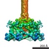 1048MC 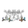 1pdfC 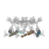 1pdiC 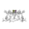 1pdjC 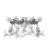 1pdmC 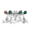 1pdpC M: map data used to model this data C: citing same article ( |
|---|---|
| Similar structure data |


-Links
 PDBj
PDBj


- Assembly
| Deposited unit | 
|
|---|---|
| 1 |
|
| Symmetry | Point symmetry: (Schoenflies symbol: C6 (6 fold cyclic)) |
-Components
| #1: Protein | Mass: 63183.723 Da / Num. of mol.: 3 / Source method: isolated from a natural source / Source: (natural) Enterobacteria phage T4 (virus) / Genus: T4-like viruses / Species: Enterobacteria phage T4 sensu lato / References: UniProt: P16009, lysozyme |
|---|
-Experimental details
-Experiment
| Experiment | Method: ELECTRON MICROSCOPY |
|---|---|
| EM experiment | Aggregation state: PARTICLE / 3D reconstruction method: single particle reconstruction |
- Sample preparation
| Component |
| |||||||||||||||
|---|---|---|---|---|---|---|---|---|---|---|---|---|---|---|---|---|
| Buffer solution | Name: water / pH: 7 / Details: water | |||||||||||||||
| Specimen | Conc.: 5 mg/ml / Embedding applied: NO / Shadowing applied: NO / Staining applied: NO / Vitrification applied: YES | |||||||||||||||
| Specimen support | Details: holey carbon | |||||||||||||||
| Vitrification | Cryogen name: ETHANE / Details: ethane vitrification | |||||||||||||||
| Crystal grow | *PLUS Method: electron microscopy |
- Electron microscopy imaging
| Microscopy | Model: FEI/PHILIPS CM300FEG/T / Date: Jan 30, 2001 |
|---|---|
| Electron gun | Electron source: FIELD EMISSION GUN / Accelerating voltage: 300 kV / Illumination mode: FLOOD BEAM |
| Electron lens | Mode: BRIGHT FIELDBright-field microscopy / Nominal magnification: 45000 X / Calibrated magnification: 47000 X / Nominal defocus max: 5000 nm / Nominal defocus min: 1200 nm / Cs: 2 mm |
| Specimen holder | Temperature: 70 K / Tilt angle max: 0 ° / Tilt angle min: 0 ° |
| Image recording | Electron dose: 25 e/Å2 / Film or detector model: KODAK SO-163 FILM |
- Processing
| EM software |
| ||||||||||||
|---|---|---|---|---|---|---|---|---|---|---|---|---|---|
| CTF correction | Details: CTF was corrected for each particle image individually | ||||||||||||
| Symmetry | Point symmetry: C6 (6 fold cyclic) | ||||||||||||
| 3D reconstruction | Method: model based projection matching / Resolution: 12 Å / Num. of particles: 945 / Nominal pixel size: 3.11 Å / Actual pixel size: 2.98 Å / Magnification calibration: TMV images Details: a modified version of SPIDER program was used for the reconstruction Symmetry type: POINT | ||||||||||||
| Atomic model building | Protocol: OTHER / Space: REAL / Target criteria: Correlation Coefficient maximization / Details: REFINEMENT PROTOCOL--Laplacian filtered real space | ||||||||||||
| Atomic model building | PDB-ID: 1K28 Accession code: 1K28 / Source name: PDB / Type: experimental model | ||||||||||||
| Refinement step | Cycle: LAST
|
