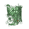
 Movie
Movie Controller


+ Open data

- Basic information
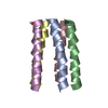
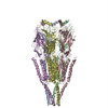
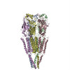
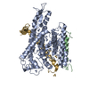
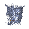
| Entry | Database: PDB / ID: 1oed | ||||||
|---|---|---|---|---|---|---|---|
| Title | STRUCTURE OF ACETYLCHOLINE RECEPTOR PORE FROM ELECTRON IMAGES | ||||||
 Components Components |
| ||||||
 Keywords Keywords | ION CHANNEL/RECEPTOR /  ION CHANNEL / TUBULAR CRYSTAL / ACETYLCHOLINE RECEPTOR / TRANSMEMBRANE / ION CHANNEL-RECEPTOR complex ION CHANNEL / TUBULAR CRYSTAL / ACETYLCHOLINE RECEPTOR / TRANSMEMBRANE / ION CHANNEL-RECEPTOR complex | ||||||
| Function / homology |  Function and homology information Function and homology informationacetylcholine-gated channel complex / acetylcholine-gated monoatomic cation-selective channel activity / acetylcholine receptor signaling pathway / transmembrane signaling receptor activity / postsynaptic membraneSimilarity search - Function | ||||||
| Biological species |  Torpedo marmorata (marbled electric ray) Torpedo marmorata (marbled electric ray) | ||||||
| Method | ELECTRON MICROSCOPY / helical reconstruction / cryo EM / Resolution: 4 Å | ||||||
 Authors Authors | Miyazawa, A. / Fujiyoshi, Y. / Unwin, N. | ||||||
 Citation Citation | Journal: Nature / Year: 2003 Title: Structure and gating mechanism of the acetylcholine receptor pore. Authors: Atsuo Miyazawa / Yoshinori Fujiyoshi / Nigel Unwin /  Abstract: The nicotinic acetylcholine receptor controls electrical signalling between nerve and muscle cells by opening and closing a gated, membrane-spanning pore. Here we present an atomic model of the ...The nicotinic acetylcholine receptor controls electrical signalling between nerve and muscle cells by opening and closing a gated, membrane-spanning pore. Here we present an atomic model of the closed pore, obtained by electron microscopy of crystalline postsynaptic membranes. The pore is shaped by an inner ring of 5 alpha-helices, which curve radially to create a tapering path for the ions, and an outer ring of 15 alpha-helices, which coil around each other and shield the inner ring from the lipids. The gate is a constricting hydrophobic girdle at the middle of the lipid bilayer, formed by weak interactions between neighbouring inner helices. When acetylcholine enters the ligand-binding domain, it triggers rotations of the protein chains on opposite sides of the entrance to the pore. These rotations are communicated through the inner helices, and open the pore by breaking the girdle apart. #1: Journal: J Mol Biol / Year: 2002 Title: Activation of the nicotinic acetylcholine receptor involves a switch in conformation of the alpha subunits. Authors: N Unwin / A Miyazawa / J Li / Y Fujiyoshi /  Abstract: The nicotinic acetylcholine (ACh) receptor belongs to a superfamily of synaptic ion channels that open in response to the binding of chemical transmitters. Their mechanism of activation is not known ...The nicotinic acetylcholine (ACh) receptor belongs to a superfamily of synaptic ion channels that open in response to the binding of chemical transmitters. Their mechanism of activation is not known in detail, but a time-resolved electron microscopic study of the muscle-type ACh receptor had suggested that a local disturbance in the ligand-binding region and consequent rotations in the ligand-binding alpha subunits, connecting to the transmembrane portion, are involved. A more precise interpretation of this structural change is given here, based on comparison of the extracellular domain of the ACh receptor with an ACh-binding protein (AChBP) to which a putative agonist is bound. We find that, to a good approximation, there are two alternative extended conformations of the ACh receptor subunits, one characteristic of either alpha subunit before activation, and the other characteristic of all three non-alpha subunits and the protomer of AChBP. Substitution in the three-dimensional maps of alpha by non-alpha subunits mimics the changes seen on activation, suggesting that the structures of the alpha subunits are modified initially by their interactions with neighbouring subunits and switch to the non-alpha form when ACh binds. This structural change, which entails 15-16 degrees rotations of the inner pore-facing parts of the alpha subunits, most likely acts as the trigger that opens the gate in the membrane-spanning pore. #2: Journal: J Mol Biol / Year: 1993 Title: Nicotinic acetylcholine receptor at 9 A resolution. Authors: N Unwin / Abstract: The nicotinic acetylcholine receptor is a cation-selective, ligand-gated ion channel, involved in signal transmission at the chemical synapse. This paper reports the three-dimensional appearance of ...The nicotinic acetylcholine receptor is a cation-selective, ligand-gated ion channel, involved in signal transmission at the chemical synapse. This paper reports the three-dimensional appearance of the channel in the closed conformation, at 9 A resolution. The structure was determined by electron microscopy of tubular crystals of Torpedo postsynaptic membranes embedded in amorphous ice. The analysis was carried out by averaging data from separate images, using helical diffraction methods. The images were recorded over a wide range of defocus (7000 to 18,800 A) so that all spacings in the object were well sampled. Tubes of only one kind ((-16.6) helical family) were processed, so that the Fourier terms could be averaged directly in reciprocal space. The three-dimensional map, obtained from 26 images, resolves some elements of secondary structure within the five protein subunits. In the synaptic part of each subunit, about 30 A from the bilayer surface, there is a group of three rods that are oriented predominantly perpendicular to the plane of the bilayer and twist around each other as in a left-handed coil. These rods presumably are alpha-helices. Two of them line the entrance to the channel, and the third is on the outside. The distinctive appearance of the alpha subunits in this region suggests that the rods may be involved in forming the binding pocket for acetylcholine. In the bilayer-spanning part of each subunit there is only one rod clearly visible, which forms the wall lining the pore, and so is assumed to be the transmembrane helix, M2. This rod does not form a straight path through the lipid bilayer, but bends, or kinks, near its mid-point, where it is closest to the axis of the pore, and tilts radially outwards on either side. It is flanked on the lipid-facing sides by a continuous rim of density, which seems likely to be composed of beta-sheet. A tentative alignment is made between the three-dimensional densities and the sequence of M2, based on correlation of the appearance of the rods with a special pattern of amino acid residues in the sequence. This alignment places the charged groups at the ends of M2 symmetrically on either side of the bilayer, and a highly conserved leucine residue (Leu251 of the alpha subunit) at the level of the kink.(ABSTRACT TRUNCATED AT 400 WORDS) #3: Journal: J Mol Biol / Year: 1999 Title: Nicotinic acetylcholine receptor at 4.6 A resolution: transverse tunnels in the channel wall. Authors: A Miyazawa / Y Fujiyoshi / M Stowell / N Unwin / Abstract: The nicotinic acetylcholine (ACh) receptor is the neurotransmitter-gated ion channel responsible for the rapid propagation of electrical signals between cells at the nerve/muscle synapse. We report ...The nicotinic acetylcholine (ACh) receptor is the neurotransmitter-gated ion channel responsible for the rapid propagation of electrical signals between cells at the nerve/muscle synapse. We report here the 4.6 A structure of this channel in the closed conformation, determined by electron microscopy of tubular crystals of Torpedo postsynaptic membranes embedded in amorphous ice. The analysis was conducted on images recorded at 4 K with a 300 kV field emission source, by combining data from four helical families of tubes (-16,6; -18,6; -15,7; -17,5), and applying three-dimensional corrections for lattice distortions. The study extends earlier work on the same specimen at 9 A resolution. Several features having functional implications now appear with better definition. The gate of the channel forms a narrow bridge, consisting of no more than one or two rings of side-chains, across the middle portion of the membrane-spanning pore. Tunnels, framed by twisted beta-sheet strands, are resolved in the extracellular wall of the channel connecting the water-filled vestibule to the putative ACh-binding pockets. A set of narrow openings through which ions can flow are resolved between alpha-helical segments forming part of the cytoplasmic wall of the channel. It is suggested that the extracellular tunnels are access routes to the binding pockets for ACh, and that the cytoplasmic openings serve as filters to exclude anions and other impermeant species from the vicinity of the pore. Both transverse pathways are likely to be important in achieving a rapid postsynaptic response. | ||||||
| History |
|
- Structure visualization
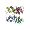
| Movie |
Movie viewer |
|---|---|
| Structure viewer | Molecule: MolmilJmol/JSmol |
- Downloads & links
-Download
| PDBx/mmCIF format | 1oed.cif.gz | 126.2 KB | Display | PDBx/mmCIF format |
|---|---|---|---|---|
| PDB format | pdb1oed.ent.gz | 95.4 KB | Display | PDB format |
| PDBx/mmJSON format | 1oed.json.gz | Tree view | PDBx/mmJSON format | |
| Others |  Other downloads Other downloads |
-Validation report
| Arichive directory | https://data.pdbj.org/pub/pdb/validation_reports/oe/1oedftp://data.pdbj.org/pub/pdb/validation_reports/oe/1oed | HTTPS FTP |
|---|
-Related structure data
| Related structure data |  1044MC M: map data used to model this data C: citing same article ( |
|---|---|
| Similar structure data |


-Links
 PDBj
PDBj

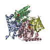
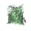
- Assembly
| Deposited unit | 
|
|---|---|
| 1 |
|
-Components
| #1: Protein | Mass: 25388.953 Da / Num. of mol.: 2 / Fragment: MEMBRANE-SPANNING DOMAIN, RESIDUES 235-461 / Source method: isolated from a natural source / Source: (natural) Torpedo marmorata (marbled electric ray) / Organ: ELECTRIC ORGAN / Tissue: DERIVED FROM MUSCLE / References: UniProt: P02711#2: Protein | | Mass: 28387.324 Da / Num. of mol.: 1 / Fragment: MEMBRANE-SPANNING DOMAIN, RESIDUES 241-490 / Source method: isolated from a natural source / Source: (natural) Torpedo marmorata (marbled electric ray) / Organ: ELECTRIC ORGAN / Tissue: DERIVED FROM MUSCLE / References: UniProt: Q6S3I0, UniProt: P02712*PLUS#3: Protein | | Mass: 29371.074 Da / Num. of mol.: 1 / Fragment: MEMBRANE-SPANNING DOMAIN, RESIDUES 246-505 / Source method: isolated from a natural source / Source: (natural) Torpedo marmorata (marbled electric ray) / Organ: ELECTRIC ORGAN / Tissue: DERIVED FROM MUSCLE / References: UniProt: Q6S3H8, UniProt: P02718*PLUS#4: Protein | | Mass: 29321.598 Da / Num. of mol.: 1 / Fragment: MEMBRANE-SPANNING DOMAIN, RESIDUES 236-495 / Source method: isolated from a natural source / Source: (natural) Torpedo marmorata (marbled electric ray) / Organ: ELECTRIC ORGAN / Tissue: DERIVED FROM MUSCLE / References: UniProt: Q6S3H9, UniProt: P02714*PLUS |
|---|
-Experimental details
-Experiment
| Experiment | Method: ELECTRON MICROSCOPY |
|---|---|
| EM experiment | Aggregation state: FILAMENT / 3D reconstruction method: helical reconstruction |
- Sample preparation
| Component | Name: POSTSYNAPTIC MEMBRANEChemical synapse / Type: VIRUS |
|---|---|
| Buffer solution | Name: SODIUM CACODYLATE / pH: 6.8 / Details: SODIUM CACODYLATE |
| Specimen | Embedding applied: NO / Shadowing applied: NO / Staining applied: NO / Vitrification applied: YES |
| Specimen support | Details: HOLEY CARBON |
| Vitrification | Instrument: HOMEMADE PLUNGER / Cryogen name: ETHANE / Details: LIQUID ETHANE |
| Crystal grow | *PLUS Method: unknown |
- Electron microscopy imaging
| Microscopy | Model: JEOL 3000SFF / Date: Oct 1, 2002 Details: THE SPECIMENS WERE TUBULAR CRYSTALS GROWN IN LOW SALT BUFFER FROM ISOLATED TORPEDO POSTSYNAPTIC MEMBRANES. THEY WERE APPLIED TO THE THE MICROSCOPE GRIDS AND FROZEN IN THE SAME SOLUTION. ...Details: THE SPECIMENS WERE TUBULAR CRYSTALS GROWN IN LOW SALT BUFFER FROM ISOLATED TORPEDO POSTSYNAPTIC MEMBRANES. THEY WERE APPLIED TO THE THE MICROSCOPE GRIDS AND FROZEN IN THE SAME SOLUTION. THESE CRYSTALS WERE TOO SMALL AND DISTORTED TO YIELD MEANINGFUL ELECTRON DIFFRACTION PATTERNS, SO BOTH THE AMPLITUDE AND THE PHASE TERMS HAD TO BE MEASURED FROM FOURIER TRANSFORMS OF THE IMAGES. THE IMAGES WERE RECORDED USING A DOSE OF 2000 ELECTRONS/NM2 DURING THE PERIOD: JAN-1996 TO OCT-2002. THE DATASETS INVOLVED 4 HELICAL FAMILIES OF TUBES, WHICH WERE ANALYSED (INCLUDING CORRECTIONS FOR DISTORTIONS) AS DESCRIBED IN THE REFERENCES. STRUCTURES WERE SYNTHESISED FROM THE AMPLITUDE AND PHASE TERMS DERIVED FROM EACH HELICAL FAMILY. THE FINAL DATASET WAS OBTAINED BY AVERAGING THESE 4 STRUCTURES IN REAL SPACE. |
|---|---|
| Electron gun | Electron source: FIELD EMISSION GUN / Accelerating voltage: 300 kV / Illumination mode: FLOOD BEAM |
| Electron lens | Mode: BRIGHT FIELDBright-field microscopy / Nominal magnification: 40000 X / Calibrated magnification: 36800 X / Nominal defocus max: 1800 nm / Nominal defocus min: 800 nm / Cs: 1.3 mm |
| Specimen holder | Temperature: 4.2 K |
| Image recording | Electron dose: 20 e/Å2 / Film or detector model: KODAK SO-163 FILM |
| Image scans | Num. digital images: 359 |
| Radiation wavelength | Relative weight: 1 |
- Processing
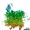
| 3D reconstruction | Resolution: 4 Å / Resolution method: FSC 0.5 CUT-OFF Details: NO REFINEMENT WAS CARRIED OUT ON THE MODEL. THE COORDINATES ARE PRELIMINARY. INCLUDED ARE ALL RESIDUES IN THE TRANSMEMBRANE SEGMENTS M1-M3 AND M4, AND THE CONNECTING LINKS M1-M2 AND M2-M3. ...Details: NO REFINEMENT WAS CARRIED OUT ON THE MODEL. THE COORDINATES ARE PRELIMINARY. INCLUDED ARE ALL RESIDUES IN THE TRANSMEMBRANE SEGMENTS M1-M3 AND M4, AND THE CONNECTING LINKS M1-M2 AND M2-M3. THE EXTENDED LOOP BETWEEN M3 AND M4, FORMING THE INTRACELLULAR DOMAIN OF THE RECEPTOR, AND SEVERAL OF THE C -TERMINAL RESIDUES ON M4 (CHAINS B, C AND E) ARE OMITTED. THE LINK BETWEEN M1 AND M2 WAS POORLY RESOLVED AND THE TRACE HERE IS ALMOST CERTAINLY WRONG IN DETAIL. IT IS HOPED THAT WITH IMPROVED DATA AND WITH REFINEMENT A MORE ACCURATE MODEL WILL BE OBTAINED. THESE COORDINATES, INCLUDING ESTIMATES OF SIDE CHAIN POSITIONS, ARE BEING MADE AVAILABLE IN THE HOPE THAT THEY WILL BE USEFUL. USERS SHOULD BEAR IN MIND THAT BECAUSE OF THE LIMITED RESOLUTION THE CONFORMATIONS OF THE SIDE CHAINS AND THEIR ATOMIC COORDINATES ARE NOT INDIVIDUALLY RELIABLE. ALSO THE ENDS OF THE HELICES ARE UNCERTAIN BY AT LEAST ONE RESIDUE. THE SURFACE LATTICE OF THE TUBULAR CRYSTALS HAS P2 SYMMETRY. THE FOUR HELICAL FAMILIES, DEFINED BY THE NUMBERS OF THE TWO PRINCIPAL HELICES REQUIRED TO FILL 360 DEGREES OF AZIMUTH, WERE: (-16,6),(-18,6),(-17,5),(-15,7). NEGATIVE INDICATES LEFT-HANDED. TO COMPARE AND COMBINE FOURIER TERMS FROM WITHIN A FAMILY IT WAS USUALLY NECESSARY TO REASSIGN LAYER-LINES TO MATCH THOSE CORRESPONDING TO A STANDARD HELICAL SELECTION RULE DEFINING A TYPICAL SET OF POSITIONS IN THE TRANSFORM. THE STANDARD SELECTION RULES WERE: L = 113N' + 314M (N = 2N') (-16,6) L = 25N' + 112M (N = 6N') (-18,6) L = 247N + 605M (-17,5) L = -91N + 662M (-15,7) WHERE L IS THE LAYER-LINE NUMBER, M IS AN INTEGER, N IS THE START NUMBER (I.E. NUMBER AROUND THE CIRCUMFERENCE) OF THE CONTRIBUTING HELIX, AND N=2N' INDICATES THAT THE START NUMBERS OCCUR IN MULTIPLES OF 2. Symmetry type: HELICAL | ||||||||||||
|---|---|---|---|---|---|---|---|---|---|---|---|---|---|
| Refinement | Highest resolution: 4 Å | ||||||||||||
| Refinement step | Cycle: LAST / Highest resolution: 4 Å
|
