 Movie
Movie Controller

[English] 日本語
 Yorodumi
- PDB-1ocy: Structure of the receptor-binding domain of the bacteriophage T4 ... -
+ Open data

- Basic information
| Entry | Database: PDB / ID: 1ocy | ||||||
|---|---|---|---|---|---|---|---|
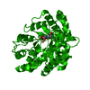
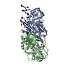
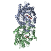
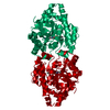
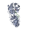
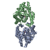
| Title | Structure of the receptor-binding domain of the bacteriophage T4 short tail fibre | ||||||
 Components Components | BACTERIOPHAGE T4 SHORT TAIL FIBRE | ||||||
 Keywords Keywords |  STRUCTURAL PROTEIN / FIBROUS PROTEIN / LIPO-POLYSACCHARIDE BINDING / BACTERIOPHAGE STRUCTURAL PROTEIN / BASEPLATE PROTEIN / GENE PRODUCT 12 STRUCTURAL PROTEIN / FIBROUS PROTEIN / LIPO-POLYSACCHARIDE BINDING / BACTERIOPHAGE STRUCTURAL PROTEIN / BASEPLATE PROTEIN / GENE PRODUCT 12 | ||||||
| Function / homology |  Function and homology information Function and homology informationvirus tail, baseplate / virus tail, fiber / entry receptor-mediated virion attachment to host cell / symbiont entry into host cell / metal ion bindingSimilarity search - Function | ||||||
| Biological species |  BACTERIOPHAGE T4 (virus) BACTERIOPHAGE T4 (virus) | ||||||
| Method | X-RAY DIFFRACTION / SYNCHROTRON / SIRAS / Resolution: 1.5 Å | ||||||
 Authors Authors | Thomassen, E. / Gielen, G. / Schuetz, M. / Miller, S. / van Raaij, M.J. | ||||||
 Citation Citation | Journal: J.Mol.Biol. / Year: 2003 Title: The Structure of the Receptor-Binding Domain of the Bacteriophage T4 Short Tail Fibre Reveals a Knitted Trimeric Metal-Binding Fold Authors: Thomassen, E. / Gielen, G. / Schuetz, M. / Schoehn, G. / Abrahams, J.P. / Miller, S. / van Raaij, M.J. #1: Journal: J.Mol.Biol. / Year: 2001 Title: Crystal Structure of a Heat- and Protease-Stable Part of the Bacteriophage T4 Short Tail Fibre Authors: van Raaij, M.J. / Schoehn, G. / Burda, M.R. / Miller, S. #2: Journal: Biol.Chem. / Year: 2001 Title: Identification and Crystallisation of a Heat- and Protease-Stable Fragment of the Bacteriophage T4 Short Tail Fibre Authors: van Raaij, M.J. / Schoehn, G. / Jaquinod, M. / Ashman, K. / Burda, M.R. / Miller, S. #3: Journal: Biol.Chem. / Year: 2000 Title: Stability of Bacteriophage T4 Short Tail Fiber Authors: Burda, M.R. / Hindennach, I. / Miller, S. #4: Journal: Eur.J.Biochem. / Year: 1999 Title: Folding of Coliphage T4 Short Tail Fiber in Vitro. Analysing the Role of a Bacteriophage-Encoded Chaperone Authors: Burda, M.R. / Miller, S. | ||||||
| History |
|
- Structure visualization
| Structure viewer | Molecule: MolmilJmol/JSmol |
|---|
- Downloads & links
-Download
| PDBx/mmCIF format | 1ocy.cif.gz | 110.5 KB | Display | PDBx/mmCIF format |
|---|---|---|---|---|
| PDB format | pdb1ocy.ent.gz | 89.9 KB | Display | PDB format |
| PDBx/mmJSON format | 1ocy.json.gz | Tree view | PDBx/mmJSON format | |
| Others |  Other downloads Other downloads |
-Validation report
| Arichive directory | https://data.pdbj.org/pub/pdb/validation_reports/oc/1ocyftp://data.pdbj.org/pub/pdb/validation_reports/oc/1ocy | HTTPS FTP |
|---|
-Related structure data
| Related structure data | |
|---|---|
| Similar structure data |


-Links
 PDBj
PDBj- Assembly
| Deposited unit | 
| |||||||||||||||||||||||||||
|---|---|---|---|---|---|---|---|---|---|---|---|---|---|---|---|---|---|---|---|---|---|---|---|---|---|---|---|---|
| 1 | 
| |||||||||||||||||||||||||||
| Unit cell |
| |||||||||||||||||||||||||||
| Components on special symmetry positions |
|
-Components
| #1: Protein | Mass: 21845.139 Da / Num. of mol.: 1 / Fragment: RECEPTOR-BINDING DOMAIN, RESIDUES 330-527 Source method: isolated from a genetically manipulated source Source: (gene. exp.) BACTERIOPHAGE T4 (virus) / Strain: DDescription: VARIANT AS PRESENT IN LABORATORY OF S. MILLER. CO-EXPRESSION WITH GP57 CHAPERONE Plasmid: PET21+ / Production host:  ESCHERICHIA COLI (E. coli) / Strain (production host): JM109 / Variant (production host): DE3 / References: UniProt: Q38160, UniProt: P10930*PLUS ESCHERICHIA COLI (E. coli) / Strain (production host): JM109 / Variant (production host): DE3 / References: UniProt: Q38160, UniProt: P10930*PLUS | ||||||
|---|---|---|---|---|---|---|---|
| #2: Chemical | ChemComp-CIT / Citric acid  Mass: 192.124 Da / Num. of mol.: 1 / Source method: obtained synthetically / Formula: C6H8O7 Mass: 192.124 Da / Num. of mol.: 1 / Source method: obtained synthetically / Formula: C6H8O7 | ||||||
| #3: Chemical | Sulfate  Mass: 96.063 Da / Num. of mol.: 2 / Source method: obtained synthetically / Formula: SO4 Mass: 96.063 Da / Num. of mol.: 2 / Source method: obtained synthetically / Formula: SO4#4: Chemical | ChemComp-ZN / |   Mass: 65.409 Da / Num. of mol.: 1 / Source method: obtained synthetically / Formula: Zn Mass: 65.409 Da / Num. of mol.: 1 / Source method: obtained synthetically / Formula: Zn#5: Water | ChemComp-HOH / | Water Mass: 18.015 Da / Num. of mol.: 516 / Source method: isolated from a natural source / Formula: H2O Mass: 18.015 Da / Num. of mol.: 516 / Source method: isolated from a natural source / Formula: H2OSequence details | T4D STRAIN VARIANT, SEQUENCE ANALYSIS OF EXPRESSION | |
-Experimental details
-Experiment
| Experiment | Method: X-RAY DIFFRACTION / Number of used crystals: 1 |
|---|
- Sample preparation
| Crystal | Density Matthews: 3.4 Å3/Da / Density % sol: 64 % | ||||||||||||||||||||||||||||||
|---|---|---|---|---|---|---|---|---|---|---|---|---|---|---|---|---|---|---|---|---|---|---|---|---|---|---|---|---|---|---|---|
| Crystal grow | pH: 5.6 Details: 15-25 % (V/V),2-METHYLPROPANE-2-OL (TERTIARY BUTANOL), 100 MM SODIUM CITRATE PH 5.6, 10% (V/V) GLYCEROL,10 MM HEPES, 150 MM SODIUM CHLORIDE | ||||||||||||||||||||||||||||||
| Crystal grow | *PLUS pH: 5.6 / Method: vapor diffusion | ||||||||||||||||||||||||||||||
| Components of the solutions | *PLUS
|
-Data collection
| Diffraction | Mean temperature: 100 K |
|---|---|
| Diffraction source | Source: SYNCHROTRON / Site: ESRF  / Beamline: ID29 / Wavelength: 0.9464 / Beamline: ID29 / Wavelength: 0.9464 |
| Detector | Type: ADSC CCD / Detector: CCD / Date: Sep 13, 2002 |
| Radiation | Protocol: SINGLE WAVELENGTH / Monochromatic (M) / Laue (L): M / Scattering type: x-ray |
| Radiation wavelength | Wavelength: 0.9464 Å / Relative weight: 1 |
| Reflection | Resolution: 1.5→15 Å / Num. obs: 96264 / % possible obs: 89.8 % / Redundancy: 6.56 % / Biso Wilson estimate: 14.103 Å2 / Rmerge(I) obs: 0.106 / Net I/σ(I): 4.122 |
| Reflection shell | Resolution: 1.5→1.58 Å / Redundancy: 3.13 % / Rmerge(I) obs: 0.28 / Mean I/σ(I) obs: 2.37 / % possible all: 53.3 |
| Reflection | *PLUS Highest resolution: 1.5 Å / Lowest resolution: 15 Å / Redundancy: 6.6 % / Rmerge(I) obs: 0.106 |
| Reflection shell | *PLUS % possible obs: 53.3 % / Redundancy: 3.1 % / Rmerge(I) obs: 0.284 |
- Processing
| Software |
| ||||||||||||||||||||
|---|---|---|---|---|---|---|---|---|---|---|---|---|---|---|---|---|---|---|---|---|---|
| Refinement | Method to determine structure: SIRAS / Resolution: 1.5→15 Å / SU B: 0.5303 / SU ML: 0.0194 / Cross valid method: THROUGHOUT / ESU R: 0.0375 / ESU R Free: 0.0354 Details: RESIDUES 85-329 COULD NOT BE MODELLED DUE TO DISORDER
| ||||||||||||||||||||
| Displacement parameters | Biso mean: 23.47 Å2
| ||||||||||||||||||||
| Refinement step | Cycle: LAST / Resolution: 1.5→15 Å
| ||||||||||||||||||||
| Refinement | *PLUS Lowest resolution: 15 Å / Rfactor Rfree: 0.152 / Rfactor Rwork: 0.143 | ||||||||||||||||||||
| Solvent computation | *PLUS | ||||||||||||||||||||
| Displacement parameters | *PLUS | ||||||||||||||||||||
| Refine LS restraints | *PLUS
| ||||||||||||||||||||
| LS refinement shell | *PLUS Highest resolution: 1.5 Å / Lowest resolution: 1.58 Å / Rfactor Rfree: 0.22 / Num. reflection Rfree: 116 / Rfactor Rwork: 0.2 / Num. reflection obs: 6339 |
