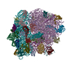
 Movie
Movie Controller


+ Open data

- Basic information
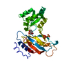
| Entry | Database: PDB / ID: 1mzp | ||||||
|---|---|---|---|---|---|---|---|
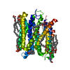
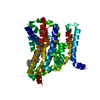
| Title | Structure of the L1 protuberance in the ribosome | ||||||
 Components Components |
| ||||||
 Keywords Keywords |  RIBOSOME / ribosomal protein / RNA-protein complex RIBOSOME / ribosomal protein / RNA-protein complex | ||||||
| Function / homology |  Function and homology informationregulation of translation / large ribosomal subunit / tRNA binding / rRNA binding / structural constituent of ribosome / translation Function and homology informationregulation of translation / large ribosomal subunit / tRNA binding / rRNA binding / structural constituent of ribosome / translationSimilarity search - Function | ||||||
| Biological species |   Sulfolobus acidocaldarius (acidophilic) Sulfolobus acidocaldarius (acidophilic) | ||||||
| Method | X-RAY DIFFRACTION / SYNCHROTRON / MAD / Resolution: 2.65 Å | ||||||
 Authors Authors | Nikulin, A. / Eliseikina, I. / Tishchenko, S. / Nevskaya, N. / Davydova, N. / Platonova, O. / Piendl, W. / Selmer, M. / Liljas, A. / Zimmermann, R. ...Nikulin, A. / Eliseikina, I. / Tishchenko, S. / Nevskaya, N. / Davydova, N. / Platonova, O. / Piendl, W. / Selmer, M. / Liljas, A. / Zimmermann, R. / Garber, M. / Nikonov, S. | ||||||
 Citation Citation | Journal: Nat.Struct.Biol. / Year: 2003 Title: Structure of the L1 protuberance in the ribosome. Authors: Nikulin, A. / Eliseikina, I. / Tishchenko, S. / Nevskaya, N. / Davydova, N. / Platonova, O. / Piendl, W. / Selmer, M. / Liljas, A. / Drygin, D. / Zimmermann, R. / Garber, M. / Nikonov, S. | ||||||
| History |
|
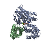
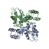
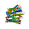
- Structure visualization
| Structure viewer | Molecule: MolmilJmol/JSmol |
|---|
- Downloads & links
-Download
| PDBx/mmCIF format | 1mzp.cif.gz | 85.3 KB | Display | PDBx/mmCIF format |
|---|---|---|---|---|
| PDB format | pdb1mzp.ent.gz | 66.8 KB | Display | PDB format |
| PDBx/mmJSON format | 1mzp.json.gz | Tree view | PDBx/mmJSON format | |
| Others |  Other downloads Other downloads |
-Validation report
| Arichive directory | https://data.pdbj.org/pub/pdb/validation_reports/mz/1mzpftp://data.pdbj.org/pub/pdb/validation_reports/mz/1mzp | HTTPS FTP |
|---|
-Related structure data
| Related structure data | |
|---|---|
| Similar structure data |


-Links
 PDBj
PDBj


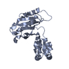
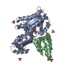
- Assembly
| Deposited unit | 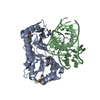
| ||||||||
|---|---|---|---|---|---|---|---|---|---|
| 1 |
| ||||||||
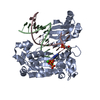
| Unit cell |
|
-Components
| #1: RNA chain | Mass: 17836.684 Da / Num. of mol.: 1 / Source method: obtained synthetically Details: Synthesis of the RNA fragment from the plasmid pDD55; sequence from cell-free(in vitro) system without living organism. | ||
|---|---|---|---|
| #2: Protein | Ribosome Mass: 24690.396 Da / Num. of mol.: 1 Source method: isolated from a genetically manipulated source Source: (gene. exp.) Sulfolobus acidocaldarius (acidophilic)Description: AUTHOR STATES TO AVOID THE POTENTIAL MISINCORPORATION OF LYSINE INSTEAD OF ARGININE, THE HOST STRAIN WAS COTRANSFORMED WITH PUBS520, A PLASMID CARRYING THE GENE FOR TRNA(ARG)AGA-AGG. Plasmid: pSacL1.4 / Production host:  Escherichia coli (E. coli) / Strain (production host): B834(DE3) / References: UniProt: P35024 Escherichia coli (E. coli) / Strain (production host): B834(DE3) / References: UniProt: P35024 | ||
| #3: Chemical |   Mass: 24.305 Da / Num. of mol.: 3 / Source method: obtained synthetically / Formula: Mg Mass: 24.305 Da / Num. of mol.: 3 / Source method: obtained synthetically / Formula: Mg#4: Water | ChemComp-HOH / | Water Mass: 18.015 Da / Num. of mol.: 94 / Source method: isolated from a natural source / Formula: H2O Mass: 18.015 Da / Num. of mol.: 94 / Source method: isolated from a natural source / Formula: H2O |
-Experimental details
-Experiment
| Experiment | Method: X-RAY DIFFRACTION / Number of used crystals: 1 |
|---|
- Sample preparation
| Crystal | Density Matthews: 3.78 Å3/Da / Density % sol: 67.2 % | ||||||||||||||||||||||||||||||||||||||||||||||||||||||||||||||||||||||||||||||||||||
|---|---|---|---|---|---|---|---|---|---|---|---|---|---|---|---|---|---|---|---|---|---|---|---|---|---|---|---|---|---|---|---|---|---|---|---|---|---|---|---|---|---|---|---|---|---|---|---|---|---|---|---|---|---|---|---|---|---|---|---|---|---|---|---|---|---|---|---|---|---|---|---|---|---|---|---|---|---|---|---|---|---|---|---|---|---|
| Crystal grow | Temperature: 277 K / Method: vapor diffusion, hanging drop / pH: 8.5 Details: PEG400, Tris HCl, KCl, MgCl2, ph 8.5, VAPOR DIFFUSION, HANGING DROP at 277K | ||||||||||||||||||||||||||||||||||||||||||||||||||||||||||||||||||||||||||||||||||||
| Components of the solutions |
| ||||||||||||||||||||||||||||||||||||||||||||||||||||||||||||||||||||||||||||||||||||
| Crystal grow | *PLUS Temperature: 4 ℃ / pH: 8.6 | ||||||||||||||||||||||||||||||||||||||||||||||||||||||||||||||||||||||||||||||||||||
| Components of the solutions | *PLUS
|
-Data collection
| Diffraction | Mean temperature: 100 K | |||||||||
|---|---|---|---|---|---|---|---|---|---|---|
| Diffraction source | Source: SYNCHROTRON / Site: EMBL/DESY, Hamburg  / Beamline: BW7A / Wavelength: 0.9762,0.9330 / Beamline: BW7A / Wavelength: 0.9762,0.9330 | |||||||||
| Detector | Type: MARRESEARCH / Detector: CCD / Date: Apr 28, 2002 | |||||||||
| Radiation | Monochromator: Double crystal focusing monochromator / Protocol: MAD / Monochromatic (M) / Laue (L): M / Scattering type: x-ray | |||||||||
| Radiation wavelength |
| |||||||||
| Reflection | Resolution: 2.65→25 Å / Num. all: 22839 / Num. obs: 22839 / % possible obs: 99.1 % / Observed criterion σ(F): 0 / Observed criterion σ(I): 0 / Redundancy: 7.8 % / Biso Wilson estimate: 70 Å2 / Rmerge(I) obs: 0.057 | |||||||||
| Reflection shell | Resolution: 2.65→2.7 Å / Rmerge(I) obs: 0.376 / % possible all: 97.2 | |||||||||
| Reflection | *PLUS Lowest resolution: 25 Å / Num. obs: 35675 / Num. measured all: 277313 | |||||||||
| Reflection shell | *PLUS % possible obs: 97.2 % / Mean I/σ(I) obs: 6 |
- Processing
| Software |
| ||||||||||||||||||||||||||||||||||||||||||||||||||||||||||||||||||||||||||||||||
|---|---|---|---|---|---|---|---|---|---|---|---|---|---|---|---|---|---|---|---|---|---|---|---|---|---|---|---|---|---|---|---|---|---|---|---|---|---|---|---|---|---|---|---|---|---|---|---|---|---|---|---|---|---|---|---|---|---|---|---|---|---|---|---|---|---|---|---|---|---|---|---|---|---|---|---|---|---|---|---|---|---|
| Refinement | Method to determine structure: MAD / Resolution: 2.65→25 Å / Rfactor Rfree error: 0.009 / Data cutoff high absF: 1263310.5 / Data cutoff high rms absF: 1263310.5 / Data cutoff low absF: 0 / Isotropic thermal model: RESTRAINED / Cross valid method: THROUGHOUT / σ(F): 2 / Stereochemistry target values: Engh & Huber
| ||||||||||||||||||||||||||||||||||||||||||||||||||||||||||||||||||||||||||||||||
| Solvent computation | Solvent model: FLAT MODEL / Bsol: 32.521 Å2 / ksol: 0.32442 e/Å3 | ||||||||||||||||||||||||||||||||||||||||||||||||||||||||||||||||||||||||||||||||
| Displacement parameters | Biso mean: 51.9 Å2
| ||||||||||||||||||||||||||||||||||||||||||||||||||||||||||||||||||||||||||||||||
| Refine analyze |
| ||||||||||||||||||||||||||||||||||||||||||||||||||||||||||||||||||||||||||||||||
| Refinement step | Cycle: LAST / Resolution: 2.65→25 Å
| ||||||||||||||||||||||||||||||||||||||||||||||||||||||||||||||||||||||||||||||||
| Refine LS restraints |
| ||||||||||||||||||||||||||||||||||||||||||||||||||||||||||||||||||||||||||||||||
| LS refinement shell | Resolution: 2.65→2.74 Å / Rfactor Rfree error: 0.043 / Total num. of bins used: 10
| ||||||||||||||||||||||||||||||||||||||||||||||||||||||||||||||||||||||||||||||||
| Xplor file |
| ||||||||||||||||||||||||||||||||||||||||||||||||||||||||||||||||||||||||||||||||
| Refinement | *PLUS Lowest resolution: 25 Å | ||||||||||||||||||||||||||||||||||||||||||||||||||||||||||||||||||||||||||||||||
| Solvent computation | *PLUS | ||||||||||||||||||||||||||||||||||||||||||||||||||||||||||||||||||||||||||||||||
| Displacement parameters | *PLUS | ||||||||||||||||||||||||||||||||||||||||||||||||||||||||||||||||||||||||||||||||
| Refine LS restraints | *PLUS
|
