 Movie
Movie Controller

[English] 日本語
 Yorodumi
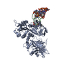
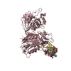
- PDB-1ma9: Crystal structure of the complex of human vitamin D binding prote... -
+ Open data

- Basic information
| Entry | Database: PDB / ID: 1ma9 | ||||||
|---|---|---|---|---|---|---|---|
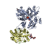
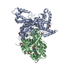
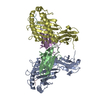
| Title | Crystal structure of the complex of human vitamin D binding protein and rabbit muscle actin | ||||||
 Components Components |
| ||||||
 Keywords Keywords | TRANSPORT PROTEIN/CONTRACTILE PROTEIN / protein-protein complex / complex formed in plasma / actin scavenger system / TRANSPORT PROTEIN-CONTRACTILE PROTEIN COMPLEX | ||||||
| Function / homology |  Function and homology information Function and homology informationvitamin transmembrane transporter activity /  calcidiol binding / vitamin transport / Vitamin D (calciferol) metabolism / vitamin D metabolic process / vitamin D binding / cytoskeletal motor activator activity / tropomyosin binding / myosin heavy chain binding / mesenchyme migration ...vitamin transmembrane transporter activity / calcidiol binding / vitamin transport / Vitamin D (calciferol) metabolism / vitamin D metabolic process / vitamin D binding / cytoskeletal motor activator activity / tropomyosin binding / myosin heavy chain binding / mesenchyme migration / troponin I binding / actin filament bundle / filamentous actin / actin filament bundle assembly / skeletal muscle thin filament assembly / striated muscle thin filament / skeletal muscle myofibril / actin monomer binding / skeletal muscle fiber development / stress fiber / titin binding / actin filament polymerization / lysosomal lumen / filopodium / actin filament / Hydrolases; Acting on acid anhydrides; Acting on acid anhydrides to facilitate cellular and subcellular movement / calcium-dependent protein binding / lamellipodium / cell body / actin binding / blood microparticle / hydrolase activity / protein domain specific binding / calcium ion binding / positive regulation of gene expression / magnesium ion binding / extracellular space / extracellular exosome / extracellular region / ATP binding / identical protein binding / cytosol / cytoplasm calcidiol binding / vitamin transport / Vitamin D (calciferol) metabolism / vitamin D metabolic process / vitamin D binding / cytoskeletal motor activator activity / tropomyosin binding / myosin heavy chain binding / mesenchyme migration ...vitamin transmembrane transporter activity / calcidiol binding / vitamin transport / Vitamin D (calciferol) metabolism / vitamin D metabolic process / vitamin D binding / cytoskeletal motor activator activity / tropomyosin binding / myosin heavy chain binding / mesenchyme migration / troponin I binding / actin filament bundle / filamentous actin / actin filament bundle assembly / skeletal muscle thin filament assembly / striated muscle thin filament / skeletal muscle myofibril / actin monomer binding / skeletal muscle fiber development / stress fiber / titin binding / actin filament polymerization / lysosomal lumen / filopodium / actin filament / Hydrolases; Acting on acid anhydrides; Acting on acid anhydrides to facilitate cellular and subcellular movement / calcium-dependent protein binding / lamellipodium / cell body / actin binding / blood microparticle / hydrolase activity / protein domain specific binding / calcium ion binding / positive regulation of gene expression / magnesium ion binding / extracellular space / extracellular exosome / extracellular region / ATP binding / identical protein binding / cytosol / cytoplasmSimilarity search - Function | ||||||
| Biological species |  Homo sapiens (human) Homo sapiens (human) Oryctolagus cuniculus (rabbit) Oryctolagus cuniculus (rabbit) | ||||||
| Method | X-RAY DIFFRACTION / SYNCHROTRON / MOLECULAR REPLACEMENT / Resolution: 2.4 Å | ||||||
 Authors Authors | Verboven, C. / Bogaerts, I. / Waelkens, E. / Rabijns, A. / Van Baelen, H. / Bouillon, R. / De Ranter, C. | ||||||
 Citation Citation | Journal: Acta Crystallogr.,Sect.D / Year: 2003 Title: Actin-DBP: the perfect structural fit? Authors: Verboven, C. / Bogaerts, I. / Waelkens, E. / Rabijns, A. / Van Baelen, H. / Bouillon, R. / De Ranter, C. #1: Journal: Acta Crystallogr.,Sect.D / Year: 2001Title: Purification, crystallization and preliminary X-ray investigation of the complex of the human vitamin D binding protein and rabbit muscle actin Authors: Bogaerts, I. / Verboven, C. / Rabijns, A. / Waelkens, E. / Van Baelen, H. / De Ranter, C. #2: Journal: Nat.Struct.Biol. / Year: 2002Title: A structural basis for the unique binding features of the human vitamin D-binding protein Authors: Verboven, C. / Rabijns, A. / De Maeyer, M. / Van Baelen, H. / Bouillon, R. / De Ranter, C. #3: Journal: Nature / Year: 1990Title: Atomic structure of the actin:DNase I complex Authors: Kabsch, W. / Mannherz, H.G. / Suck, D. / Pai, E.F. / Holmes, K.C. | ||||||
| History |
|
- Structure visualization
| Structure viewer | Molecule: MolmilJmol/JSmol |
|---|
- Downloads & links
-Download
| PDBx/mmCIF format | 1ma9.cif.gz | 169.6 KB | Display | PDBx/mmCIF format |
|---|---|---|---|---|
| PDB format | pdb1ma9.ent.gz | 138.1 KB | Display | PDB format |
| PDBx/mmJSON format | 1ma9.json.gz | Tree view | PDBx/mmJSON format | |
| Others |  Other downloads Other downloads |
-Validation report
| Arichive directory | https://data.pdbj.org/pub/pdb/validation_reports/ma/1ma9ftp://data.pdbj.org/pub/pdb/validation_reports/ma/1ma9 | HTTPS FTP |
|---|
-Related structure data


-Links
 PDBj
PDBj


- Assembly
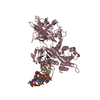
| Deposited unit | 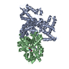
| ||||||||
|---|---|---|---|---|---|---|---|---|---|
| 1 |
| ||||||||
| Unit cell |
|
-Components
| #1: Protein | Mass: 51291.316 Da / Num. of mol.: 1 / Source method: isolated from a natural source / Details: serum / Source: (natural) Homo sapiens (human) / References: UniProt: P02774 |
|---|---|
| #2: Protein | / Alpha-actin 1 Mass: 41875.633 Da / Num. of mol.: 1 / Source method: isolated from a natural source / Details: muscle / Source: (natural) Oryctolagus cuniculus (rabbit) / References: UniProt: P68135 |
| #3: Chemical | ChemComp-MG /   Mass: 24.305 Da / Num. of mol.: 1 / Source method: obtained synthetically / Formula: Mg Mass: 24.305 Da / Num. of mol.: 1 / Source method: obtained synthetically / Formula: Mg |
| #4: Chemical | ChemComp-ATP / Adenosine triphosphate  Mass: 507.181 Da / Num. of mol.: 1 / Source method: obtained synthetically / Formula: C10H16N5O13P3 / Comment: ATP, energy-carrying molecule*YM Mass: 507.181 Da / Num. of mol.: 1 / Source method: obtained synthetically / Formula: C10H16N5O13P3 / Comment: ATP, energy-carrying molecule*YM |
| #5: Water | ChemComp-HOH / Water Mass: 18.015 Da / Num. of mol.: 258 / Source method: isolated from a natural source / Formula: H2O Mass: 18.015 Da / Num. of mol.: 258 / Source method: isolated from a natural source / Formula: H2O |
-Experimental details
-Experiment
| Experiment | Method: X-RAY DIFFRACTION / Number of used crystals: 1 |
|---|
- Sample preparation
| Crystal | Density Matthews: 2.47 Å3/Da / Density % sol: 50 % |
|---|---|
| Crystal grow | Temperature: 277 K / Method: vapor diffusion, hanging drop / pH: 6.3 Details: PEG 8000, magnesium acetate, sodium cacodylate, glycerol, pH 6.3, VAPOR DIFFUSION, HANGING DROP, temperature 277K |
| Crystal grow | *PLUS Details: Verboven, C.C., (1995) J. Steroid Biochem. Mol. Biol., 54, 11. |
-Data collection
| Diffraction | Mean temperature: 100 K |
|---|---|
| Diffraction source | Source: SYNCHROTRON / Site: EMBL/DESY, Hamburg  / Beamline: BW7B / Wavelength: 0.8423 Å / Beamline: BW7B / Wavelength: 0.8423 Å |
| Detector | Type: MARRESEARCH / Detector: IMAGE PLATE / Date: Sep 26, 2000 / Details: premirror, triangular monochromator, bent mirror |
| Radiation | Monochromator: triangular monochromator / Protocol: SINGLE WAVELENGTH / Monochromatic (M) / Laue (L): M / Scattering type: x-ray |
| Radiation wavelength | Wavelength: 0.8423 Å / Relative weight: 1 |
| Reflection | Resolution: 2.4→20 Å / Num. all: 35664 / Num. obs: 33202 / % possible obs: 93.3 % / Observed criterion σ(F): 0 / Observed criterion σ(I): -3 / Redundancy: 2.9 % / Biso Wilson estimate: 36.3 Å2 / Rmerge(I) obs: 0.042 / Rsym value: 0.042 / Net I/σ(I): 12.2 |
| Reflection shell | Resolution: 2.4→2.44 Å / Redundancy: 2.6 % / Rmerge(I) obs: 0.25 / Mean I/σ(I) obs: 1.8 / Num. unique all: 1633 / Rsym value: 0.25 / % possible all: 93.9 |
| Reflection | *PLUS Lowest resolution: 20 Å / Num. obs: 33252 / Num. measured all: 97747 |
| Reflection shell | *PLUS % possible obs: 93.9 % / Rmerge(I) obs: 0.25 |
- Processing
| Software |
| ||||||||||||||||||||||||||||||||||||
|---|---|---|---|---|---|---|---|---|---|---|---|---|---|---|---|---|---|---|---|---|---|---|---|---|---|---|---|---|---|---|---|---|---|---|---|---|---|
| Refinement | Method to determine structure: MOLECULAR REPLACEMENT Starting model: PDB ENTRIES 1ATN, 1J78 Resolution: 2.4→19.91 Å / Rfactor Rfree error: 0.005 / Data cutoff high absF: 818801.14 / Data cutoff high rms absF: 818801.14 / Data cutoff low absF: 0 / Isotropic thermal model: RESTRAINED / Cross valid method: THROUGHOUT / σ(F): 0 / Stereochemistry target values: Engh & Huber Details: simulated annealing, torsion angle dynamics, refinement target : maximum likelihood target using amplitudes
| ||||||||||||||||||||||||||||||||||||
| Solvent computation | Solvent model: FLAT MODEL / Bsol: 44.3355 Å2 / ksol: 0.336072 e/Å3 | ||||||||||||||||||||||||||||||||||||
| Displacement parameters | Biso mean: 52.2 Å2
| ||||||||||||||||||||||||||||||||||||
| Refine analyze |
| ||||||||||||||||||||||||||||||||||||
| Refinement step | Cycle: LAST / Resolution: 2.4→19.91 Å
| ||||||||||||||||||||||||||||||||||||
| Refine LS restraints |
| ||||||||||||||||||||||||||||||||||||
| LS refinement shell | Resolution: 2.4→2.55 Å / Rfactor Rfree error: 0.017 / Total num. of bins used: 6
| ||||||||||||||||||||||||||||||||||||
| Xplor file |
| ||||||||||||||||||||||||||||||||||||
| Refinement | *PLUS Rfactor Rfree: 0.2532 / Rfactor Rwork: 0.1983 | ||||||||||||||||||||||||||||||||||||
| Solvent computation | *PLUS | ||||||||||||||||||||||||||||||||||||
| Displacement parameters | *PLUS | ||||||||||||||||||||||||||||||||||||
| Refine LS restraints | *PLUS
|
