 Movie
Movie Controller

[English] 日本語
 Yorodumi
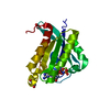
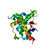
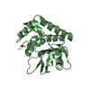
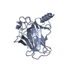
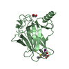
- PDB-1kxf: SINDBIS VIRUS CAPSID, (WILD-TYPE) RESIDUES 1-264, TETRAGONAL CRYS... -
+ Open data

- Basic information
| Entry | Database: PDB / ID: 1kxf | ||||||
|---|---|---|---|---|---|---|---|
| Title | SINDBIS VIRUS CAPSID, (WILD-TYPE) RESIDUES 1-264, TETRAGONAL CRYSTAL FORM (FORM II) | ||||||
 Components Components | SINDBIS VIRUS CAPSID PROTEIN | ||||||
 Keywords Keywords |  VIRAL PROTEIN / SINDBIS VIRUS CAPSID PROTEIN / CHYMOTRYPSIN-LIKE SERINE PROTEINASE / WILD TYPE / VIRUS CAPSID PROTEIN VIRAL PROTEIN / SINDBIS VIRUS CAPSID PROTEIN / CHYMOTRYPSIN-LIKE SERINE PROTEINASE / WILD TYPE / VIRUS CAPSID PROTEIN | ||||||
| Function / homology |  Function and homology information Function and homology informationicosahedral viral capsid, spike / togavirin / T=4 icosahedral viral capsid / ubiquitin-like protein ligase binding / symbiont-mediated suppression of host toll-like receptor signaling pathway / clathrin-dependent endocytosis of virus by host cell / host cell cytoplasm / membrane fusion / serine-type endopeptidase activity / fusion of virus membrane with host endosome membrane ...icosahedral viral capsid, spike / togavirin / T=4 icosahedral viral capsid / ubiquitin-like protein ligase binding / symbiont-mediated suppression of host toll-like receptor signaling pathway / clathrin-dependent endocytosis of virus by host cell / host cell cytoplasm / membrane fusion / serine-type endopeptidase activity / fusion of virus membrane with host endosome membrane / viral envelope / host cell nucleus / structural molecule activity / virion attachment to host cell / host cell plasma membrane / virion membrane / proteolysis / RNA binding / membraneSimilarity search - Function | ||||||
| Biological species |  Sindbis virus Sindbis virus | ||||||
| Method | X-RAY DIFFRACTION / Resolution: 2.38 Å | ||||||
 Authors Authors | Choi, H.-K. / Rossmann, M.G. | ||||||
 Citation Citation | Journal: J.Mol.Biol. / Year: 1996 Title: Structural analysis of Sindbis virus capsid mutants involving assembly and catalysis. Authors: Choi, H.K. / Lee, S. / Zhang, Y.P. / McKinney, B.R. / Wengler, G. / Rossmann, M.G. / Kuhn, R.J. #1: Journal: J.Mol.Biol. / Year: 1993Title: Refined Structure of Sindbis Virus Core Protein and Comparison with Other Chymotrypsin-Like Serine Proteinase Structures Authors: Tong, L. / Wengler, G. / Rossmann, M.G. #2: Journal: Acta Crystallogr.,Sect.A / Year: 1992Title: The Structure Determination of Sindbis Virus Core Protein Using Isomorphous Replacement and Molecular Replacement Averaging between Two Crystal Forms Authors: Tong, L. / Choi, H.-K. / Minor, W. / Rossmann, M.G. #3: Journal: Nature / Year: 1991Title: Structure of Sindbis Virus Core Protein Reveals a Chymotrypsin-Like Serine Proteinase and the Organization of the Virion Authors: Choi, H.K. / Tong, L. / Minor, W. / Dumas, P. / Boege, U. / Rossmann, M.G. / Wengler, G. | ||||||
| History |
|
- Structure visualization
| Structure viewer | Molecule: MolmilJmol/JSmol |
|---|
- Downloads & links
-Download
| PDBx/mmCIF format | 1kxf.cif.gz | 41.4 KB | Display | PDBx/mmCIF format |
|---|---|---|---|---|
| PDB format | pdb1kxf.ent.gz | 29.1 KB | Display | PDB format |
| PDBx/mmJSON format | 1kxf.json.gz | Tree view | PDBx/mmJSON format | |
| Others |  Other downloads Other downloads |
-Validation report
| Arichive directory | https://data.pdbj.org/pub/pdb/validation_reports/kx/1kxfftp://data.pdbj.org/pub/pdb/validation_reports/kx/1kxf | HTTPS FTP |
|---|
-Related structure data
| Related structure data | 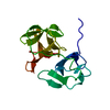 1kxaC  1kxbC  1kxcC  1kxdC  1kxeC  2snwC C: citing same article ( |
|---|---|
| Similar structure data |


-Links
 PDBj
PDBj


- Assembly
| Deposited unit | 
| ||||||||
|---|---|---|---|---|---|---|---|---|---|
| 1 |
| ||||||||
| Unit cell |
|
-Components
| #1: Protein | Mass: 17418.680 Da / Num. of mol.: 1 / Fragment: MET 1 - ALA 264 OF THE NATIVE SINDBIS CAPSID Source method: isolated from a genetically manipulated source Source: (gene. exp.) Sindbis virus / Genus: Alphavirus / Organ: KIDNEY / Production host:  Cricetinae (hamsters) Cricetinae (hamsters)References: UniProt: P03316, Hydrolases; Acting on peptide bonds (peptidases); Serine endopeptidases |
|---|---|
| #2: Water | ChemComp-HOH / Water Mass: 18.015 Da / Num. of mol.: 16 / Source method: isolated from a natural source / Formula: H2O Mass: 18.015 Da / Num. of mol.: 16 / Source method: isolated from a natural source / Formula: H2O |
| Sequence details | crystallized construct 1-264 was possibly cleaved during crystallization somewhere before residue ...crystallized construct 1-264 was possibly cleaved during crystallization somewhere before residue 106. The entry was annotated as 106-264 fragment |
-Experimental details
-Experiment
| Experiment | Method: X-RAY DIFFRACTION |
|---|
- Sample preparation
| Crystal | Density Matthews: 2.56 Å3/Da / Density % sol: 51.95 % | ||||||||||||||||||||
|---|---|---|---|---|---|---|---|---|---|---|---|---|---|---|---|---|---|---|---|---|---|
| Crystal grow | *PLUS Method: vapor diffusion, hanging drop / PH range low: 8.5 / PH range high: 7.6 | ||||||||||||||||||||
| Components of the solutions | *PLUS
|
-Data collection
| Diffraction source | Wavelength: 1.5418 |
|---|---|
| Detector | Type: SIEMENS / Detector: AREA DETECTOR / Date: Feb 26, 1992 |
| Radiation | Monochromatic (M) / Laue (L): M / Scattering type: x-ray |
| Radiation wavelength | Wavelength: 1.5418 Å / Relative weight: 1 |
| Reflection | Highest resolution: 2.38 Å / Num. obs: 7908 / % possible obs: 96.2 % / Observed criterion σ(I): 1 / Redundancy: 2.42 % / Rmerge(I) obs: 0.042 |
| Reflection | *PLUS Highest resolution: 2.4 Å / Lowest resolution: 9999 Å / Num. measured all: 19176 |
| Reflection shell | *PLUS Highest resolution: 2.4 Å / Lowest resolution: 2.6 Å / % possible obs: 90.4 % / Rmerge(I) obs: 0.134 |
- Processing
| Software |
| ||||||||||||||||||||||||||||||||||||||||||||||||||||||||||||
|---|---|---|---|---|---|---|---|---|---|---|---|---|---|---|---|---|---|---|---|---|---|---|---|---|---|---|---|---|---|---|---|---|---|---|---|---|---|---|---|---|---|---|---|---|---|---|---|---|---|---|---|---|---|---|---|---|---|---|---|---|---|
| Refinement | Resolution: 2.38→6 Å / σ(F): 2
| ||||||||||||||||||||||||||||||||||||||||||||||||||||||||||||
| Displacement parameters | Biso mean: 28.4 Å2 | ||||||||||||||||||||||||||||||||||||||||||||||||||||||||||||
| Refinement step | Cycle: LAST / Resolution: 2.38→6 Å
| ||||||||||||||||||||||||||||||||||||||||||||||||||||||||||||
| Refine LS restraints |
| ||||||||||||||||||||||||||||||||||||||||||||||||||||||||||||
| Software | *PLUS Name: X-PLOR / Classification: refinement | ||||||||||||||||||||||||||||||||||||||||||||||||||||||||||||
| Refinement | *PLUS Highest resolution: 2.4 Å | ||||||||||||||||||||||||||||||||||||||||||||||||||||||||||||
| Solvent computation | *PLUS | ||||||||||||||||||||||||||||||||||||||||||||||||||||||||||||
| Displacement parameters | *PLUS | ||||||||||||||||||||||||||||||||||||||||||||||||||||||||||||
| Refine LS restraints | *PLUS
|
