 Movie
Movie Controller


+ Open data

- Basic information
| Entry | Database: PDB / ID: 1ijg | ||||||
|---|---|---|---|---|---|---|---|
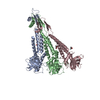
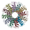
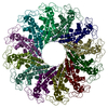
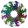
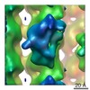
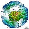
| Title | Structure of the Bacteriophage phi29 Head-Tail Connector Protein | ||||||
 Components Components | UPPER COLLAR PROTEIN | ||||||
 Keywords Keywords |  VIRAL PROTEIN / alpha-helical bundle / RNA binding motif VIRAL PROTEIN / alpha-helical bundle / RNA binding motif | ||||||
| Function / homology |  Function and homology information Function and homology informationviral portal complex / viral procapsid / symbiont genome ejection through host cell envelope, short tail mechanism / viral DNA genome packaging / RNA bindingSimilarity search - Function | ||||||
| Biological species |   Bacillus phage phi29 (virus) Bacillus phage phi29 (virus) | ||||||
| Method | X-RAY DIFFRACTION / SYNCHROTRON / MOLECULAR REPLACEMENT / Resolution: 2.9 Å | ||||||
 Authors Authors | Simpson, A.A. / Leiman, P.G. / Tao, Y. / He, Y. / Badasso, M. / Jardine, P.J. / Anderson, D.L. / Rossmann, M.G. | ||||||
 Citation Citation | Journal: Acta Crystallogr.,Sect.D / Year: 2001 Title: Structure determination of the head-tail connector of bacteriophage phi29. Authors: Simpson, A.A. / Leiman, P.G. / Tao, Y. / He, Y. / Badasso, M.O. / Jardine, P.J. / Anderson, D.L. / Rossmann, M.G. | ||||||
| History |
|
- Structure visualization
| Structure viewer | Molecule: MolmilJmol/JSmol |
|---|
- Downloads & links
-Download
| PDBx/mmCIF format | 1ijg.cif.gz | 628.7 KB | Display | PDBx/mmCIF format |
|---|---|---|---|---|
| PDB format | pdb1ijg.ent.gz | 523.4 KB | Display | PDB format |
| PDBx/mmJSON format | 1ijg.json.gz | Tree view | PDBx/mmJSON format | |
| Others |  Other downloads Other downloads |
-Validation report
| Arichive directory | https://data.pdbj.org/pub/pdb/validation_reports/ij/1ijgftp://data.pdbj.org/pub/pdb/validation_reports/ij/1ijg | HTTPS FTP |
|---|
-Related structure data
| Related structure data | 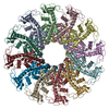 1jnbC  1fouS S: Starting model for refinement C: citing same article ( |
|---|---|
| Similar structure data |


-Links
 PDBj
PDBj- Assembly
| Deposited unit | 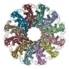
| ||||||||
|---|---|---|---|---|---|---|---|---|---|
| 1 |
| ||||||||
| Unit cell |
| ||||||||
| Details | The biological assembly is a dodecamer luckily comprising the asymmetric unit. |
-Components
| #1: Protein | Mass: 35917.293 Da / Num. of mol.: 12 Source method: isolated from a genetically manipulated source Source: (gene. exp.) Bacillus phage phi29 (virus) / Genus: Phi29-like viruses / Gene: 10 / Production host:  Escherichia coli (E. coli) / References: UniProt: P04332 Escherichia coli (E. coli) / References: UniProt: P04332#2: Water | ChemComp-HOH / | Water Mass: 18.015 Da / Num. of mol.: 753 / Source method: isolated from a natural source / Formula: H2O Mass: 18.015 Da / Num. of mol.: 753 / Source method: isolated from a natural source / Formula: H2O |
|---|
-Experimental details
-Experiment
| Experiment | Method: X-RAY DIFFRACTION / Number of used crystals: 1 |
|---|
- Sample preparation
| Crystal | Density Matthews: 3.56 Å3/Da / Density % sol: 65.21 % | ||||||||||||||||||||||||
|---|---|---|---|---|---|---|---|---|---|---|---|---|---|---|---|---|---|---|---|---|---|---|---|---|---|
| Crystal grow | Temperature: 298 K / Method: vapor diffusion, hanging drop / pH: 8 Details: MPD, calcium cloride, tris buffer, pH 8.0, VAPOR DIFFUSION, HANGING DROP, temperature 298.0K | ||||||||||||||||||||||||
| Crystal grow | *PLUS Method: unknown | ||||||||||||||||||||||||
| Components of the solutions | *PLUS
|
-Data collection
| Diffraction | Mean temperature: 100 K |
|---|---|
| Diffraction source | Source: SYNCHROTRON / Site: APS  / Beamline: 14-BM-C / Wavelength: 1 Å / Beamline: 14-BM-C / Wavelength: 1 Å |
| Detector | Type: ADSC QUANTUM 4 / Detector: CCD / Date: Aug 15, 1999 / Details: bent conical Si-mirror (Rh coating) |
| Radiation | Monochromator: bend cylindrical Ge(111) monochromator / Protocol: SINGLE WAVELENGTH / Monochromatic (M) / Laue (L): M / Scattering type: x-ray |
| Radiation wavelength | Wavelength: 1 Å / Relative weight: 1 |
| Reflection | Resolution: 2.9→50 Å / Num. all: 112772 / Num. obs: 110311 / % possible obs: 97.8 % / Observed criterion σ(F): 1 / Observed criterion σ(I): 1 / Redundancy: 3.4 % / Biso Wilson estimate: 58.4 Å2 / Rmerge(I) obs: 0.088 / Net I/σ(I): 28.9 |
| Reflection shell | Resolution: 2.9→2.92 Å / Redundancy: 3.2 % / Rmerge(I) obs: 0.333 / % possible all: 99.6 |
| Reflection | *PLUS Redundancy: 3.54 % |
| Reflection shell | *PLUS % possible obs: 99 % |
- Processing
| Software |
| |||||||||||||||||||||||||
|---|---|---|---|---|---|---|---|---|---|---|---|---|---|---|---|---|---|---|---|---|---|---|---|---|---|---|
| Refinement | Method to determine structure: MOLECULAR REPLACEMENT Starting model: PDB ENTRY 1FOU Resolution: 2.9→500 Å / Isotropic thermal model: anisotropic / Cross valid method: THROUGHOUT / σ(F): 0 / σ(I): 0 / Stereochemistry target values: Engh & Huber
| |||||||||||||||||||||||||
| Displacement parameters | Biso mean: 54.2 Å2
| |||||||||||||||||||||||||
| Refine analyze | Luzzati coordinate error obs: 0.363 Å | |||||||||||||||||||||||||
| Refinement step | Cycle: LAST / Resolution: 2.9→500 Å
| |||||||||||||||||||||||||
| Refine LS restraints |
| |||||||||||||||||||||||||
| LS refinement shell | Resolution: 2.9→2.92 Å / Total num. of bins used: 50
| |||||||||||||||||||||||||
| Software | *PLUS Name: CNS / Version: 1 / Classification: refinement | |||||||||||||||||||||||||
| Refinement | *PLUS Highest resolution: 2.9 Å / Lowest resolution: 500 Å / σ(F): 0 / % reflection Rfree: 2.27 % | |||||||||||||||||||||||||
| Solvent computation | *PLUS | |||||||||||||||||||||||||
| Displacement parameters | *PLUS Biso mean: 54.2 Å2 | |||||||||||||||||||||||||
| LS refinement shell | *PLUS Rfactor Rfree: 0.29 / % reflection Rfree: 2.5 % / Rfactor Rwork: 0.324 |
