[English] 日本語
Yorodumi
- PDB-1g1x: STRUCTURE OF RIBOSOMAL PROTEINS S15, S6, S18, AND 16S RIBOSOMAL RNA -

+
Open data


ID or keywords:

Loading...

-
Basic information

Entry
Database: PDB / ID: 1g1x
TitleSTRUCTURE OF RIBOSOMAL PROTEINS S15, S6, S18, AND 16S RIBOSOMAL RNA
Components
  • (16S RIBOSOMAL RNA) x 2
  • 30S RIBOSOMAL PROTEIN S15
  • 30S RIBOSOMAL PROTEIN S18
  • 30S RIBOSOMAL PROTEIN S6
KeywordsRIBOSOME / ribosomal proteins S15 / S6 / S18 / S30 ribosomal subunit / RNA
Function / homology
Function and homology information


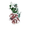
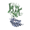
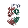
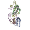
rRNA binding / ribosome / structural constituent of ribosome / translation / ribonucleoprotein complex / cytoplasm
Similarity search - Function
Ribosomal protein S18 / 30s Ribosomal Protein S18 / Ribosomal protein S6/Translation elongation factor EF1B / S15/NS1, RNA-binding / Ribosomal protein S6, conserved site / Ribosomal protein S6 signature. / Ribosomal protein S6, plastid/chloroplast / Ribosomal protein S18, conserved site / Ribosomal protein S18 signature. / Ribosomal protein S15, bacterial-type ...Ribosomal protein S18 / 30s Ribosomal Protein S18 / Ribosomal protein S6/Translation elongation factor EF1B / S15/NS1, RNA-binding / Ribosomal protein S6, conserved site / Ribosomal protein S6 signature. / Ribosomal protein S6, plastid/chloroplast / Ribosomal protein S18, conserved site / Ribosomal protein S18 signature. / Ribosomal protein S15, bacterial-type / Ribosomal protein S6 / Ribosomal protein S6 / Ribosomal protein S6 superfamily / Translation elongation factor EF1B/ribosomal protein S6 / Ribosomal protein S18 / Ribosomal protein S18 / Ribosomal protein S18 superfamily / Few Secondary Structures / Irregular / Ribosomal_S15 / Ribosomal protein S15 signature. / Ribosomal protein S15 / Ribosomal protein S15 / S15/NS1, RNA-binding / Helix Hairpins / Alpha-Beta Plaits / 2-Layer Sandwich / Orthogonal Bundle / Mainly Alpha / Alpha Beta
Similarity search - Domain/homology
RNA / RNA (> 10) / Small ribosomal subunit protein uS15 / Small ribosomal subunit protein bS6 / Small ribosomal subunit protein bS18
Similarity search - Component
Biological speciesThermus thermophilus (bacteria)
MethodX-RAY DIFFRACTION / SYNCHROTRON / MIR / Resolution: 2.6 Å
AuthorsAgalarov, S.C. / Prasad, G.S. / Funke, P.M. / Stout, C.D. / Williamson, J.R.
CitationJournal: Science / Year: 2000
Title: Structure of the S15,S6,S18-rRNA complex: assembly of the 30S ribosome central domain.
Authors: Agalarov, S.C. / Sridhar Prasad, G. / Funke, P.M. / Stout, C.D. / Williamson, J.R.
History
DepositionOct 13, 2000Deposition site: RCSB / Processing site: RCSB
SupersessionOct 30, 2000ID: 1EKC
Revision 1.0Oct 30, 2000Provider: repository / Type: Initial release
Revision 1.1Apr 27, 2008Group: Version format compliance
Revision 1.2Jul 13, 2011Group: Version format compliance
Revision 1.3Feb 7, 2024Group: Data collection / Database references
Category: chem_comp_atom / chem_comp_bond ...chem_comp_atom / chem_comp_bond / database_2 / struct_ref_seq_dif
Item: _database_2.pdbx_DOI / _database_2.pdbx_database_accession / _struct_ref_seq_dif.details

-
Structure visualization

Structure viewerMolecule:
MolmilJmol/JSmol

Downloads & links

-
Assembly

Deposited unit
D: 16S RIBOSOMAL RNA
E: 16S RIBOSOMAL RNA
I: 16S RIBOSOMAL RNA
J: 16S RIBOSOMAL RNA
A: 30S RIBOSOMAL PROTEIN S6
B: 30S RIBOSOMAL PROTEIN S15
C: 30S RIBOSOMAL PROTEIN S18
F: 30S RIBOSOMAL PROTEIN S6
G: 30S RIBOSOMAL PROTEIN S15
H: 30S RIBOSOMAL PROTEIN S18


Theoretical massNumber of molelcules
Total (without water)120,29310
Polymers120,29310
Non-polymers00
Water0
1
D: 16S RIBOSOMAL RNA
E: 16S RIBOSOMAL RNA
A: 30S RIBOSOMAL PROTEIN S6
B: 30S RIBOSOMAL PROTEIN S15
C: 30S RIBOSOMAL PROTEIN S18


Theoretical massNumber of molelcules
Total (without water)60,1475
Polymers60,1475
Non-polymers00
Water0
TypeNameSymmetry operationNumber
identity operation1_555x,y,z1
2
I: 16S RIBOSOMAL RNA
J: 16S RIBOSOMAL RNA
F: 30S RIBOSOMAL PROTEIN S6
G: 30S RIBOSOMAL PROTEIN S15
H: 30S RIBOSOMAL PROTEIN S18


Theoretical massNumber of molelcules
Total (without water)60,1475
Polymers60,1475
Non-polymers00
Water0
TypeNameSymmetry operationNumber
identity operation1_555x,y,z1
Unit cell
Length a, b, c (Å)169.5, 169.5, 113.8
Angle α, β, γ (deg.)90.0, 90.0, 120.0
Int Tables number170
Cell settinghexagonal
Space group name H-MP65

-
Components

#1: RNA chain 16S RIBOSOMAL RNA /


Mass: 13576.196 Da / Num. of mol.: 2 / Fragment: RESIDUES 582-675 / Source method: obtained synthetically
#2: RNA chain 16S RIBOSOMAL RNA /


Mass: 14102.433 Da / Num. of mol.: 2 / Fragment: RESIDUES 716-759 / Source method: obtained synthetically
#3: Protein 30S RIBOSOMAL PROTEIN S6 /


Mass: 11732.494 Da / Num. of mol.: 2 / Source method: isolated from a natural source / Source: (natural) Thermus thermophilus (bacteria) / References: UniProt: Q5SLP8
#4: Protein 30S RIBOSOMAL PROTEIN S15 /


Mass: 10491.223 Da / Num. of mol.: 2 / Source method: isolated from a natural source / Source: (natural) Thermus thermophilus (bacteria) / References: UniProt: Q5SJ76
#5: Protein 30S RIBOSOMAL PROTEIN S18 /


Mass: 10244.272 Da / Num. of mol.: 2 / Source method: isolated from a natural source / Source: (natural) Thermus thermophilus (bacteria) / References: UniProt: Q5SLQ0

-
Experimental details

-
Experiment

ExperimentMethod: X-RAY DIFFRACTION / Number of used crystals: 1

-
Sample preparation

CrystalDensity Matthews: 3.4 Å3/Da / Density % sol: 75 %
Crystal growTemperature: 293 K / Method: vapor diffusion, sitting drop / pH: 7
Details: 1.8 M (NH4)2SO4, 20 mM MgCl2, 50 mM Potassium Cacodylate, pH 7.0, VAPOR DIFFUSION, SITTING DROP, temperature 293K
Components of the solutions
IDNameCrystal-IDSol-ID
1MgCl211
2Potassium Cacodylate11
3(NH4)2SO411
4(NH4)2SO412
Crystal grow
*PLUS
pH: 6.5
Components of the solutions
*PLUS
IDConc.Common nameCrystal-IDSol-IDChemical formula
11.8 Mammonium sulfate1reservoir
220 mM1reservoirMgCl2
350 mMpotassium cacodylate1reservoir
41

-
Data collection

DiffractionMean temperature: 100 K
Diffraction sourceSource: SYNCHROTRON / Site: SSRL / Beamline: BL9-1 / Wavelength: 0.98
DetectorType: MARRESEARCH / Detector: IMAGE PLATE / Date: Apr 26, 1999
RadiationProtocol: SINGLE WAVELENGTH / Monochromatic (M) / Laue (L): M / Scattering type: x-ray
Radiation wavelengthWavelength: 0.98 Å / Relative weight: 1
ReflectionResolution: 2.6→50 Å / Num. obs: 57348 / % possible obs: 99.9 % / Observed criterion σ(I): 0 / Redundancy: 3.8 % / Biso Wilson estimate: 71.4 Å2 / Rmerge(I) obs: 0.051 / Rsym value: 0.279 / Net I/σ(I): 9.9
Reflection shellResolution: 2.6→2.69 Å / % possible obs: 99.9 % / Redundancy: 3.8 % / Rmerge(I) obs: 0.41 / Mean I/σ(I) obs: 1.7 / Rsym value: 0.279 / % possible all: 99.9
Reflection
*PLUS
Num. measured all: 264392

-
Processing

Software
NameVersionClassification
MLPHAREphasing
X-PLORrefinement
MOSFLMdata reduction
CCP4(TRUNCATE)data scaling
RefinementMethod to determine structure: MIR / Resolution: 2.6→50 Å / Cross valid method: THROUGHOUT / σ(F): 0
RfactorNum. reflection% reflectionSelection details
Rfree0.298 1289 2.2 %random
Rwork0.266 ---
obs-57348 99.9 %-
Displacement parametersBiso mean: 44.48 Å2
Refine analyze
FreeObs
Luzzati coordinate error0.68 Å0.58 Å
Luzzati d res low-5 Å
Luzzati sigma a0.65 Å0.73 Å
Refinement stepCycle: LAST / Resolution: 2.6→50 Å
ProteinNucleic acidLigandSolventTotal
Num. atoms3897 3646 0 0 7543
Refine LS restraints
Refine-IDTypeDev ideal
X-RAY DIFFRACTIONx_bond_d0.023
X-RAY DIFFRACTIONx_angle_deg2.22
X-RAY DIFFRACTIONx_dihedral_angle_d19.45
X-RAY DIFFRACTIONx_improper_angle_d1.81
X-RAY DIFFRACTIONx_mcbond_it4.34
X-RAY DIFFRACTIONx_mcangle_it6.35
X-RAY DIFFRACTIONx_scbond_it5.58
X-RAY DIFFRACTIONx_scangle_it7.75
LS refinement shellResolution: 2.6→2.72 Å / Total num. of bins used: 8
RfactorNum. reflection% reflection
Rfree0.48 60 2.2 %
Rwork0.42 7071 -
obs--99.9 %
Xplor file
Refine-IDSerial noParam fileTopol file
X-RAY DIFFRACTION1protein_rep.paramprotein.top
X-RAY DIFFRACTION2dna-rna_rep.paramdna-rna.top
Software
*PLUS
Name: X-PLOR / Classification: refinement
Refinement
*PLUS
Highest resolution: 2.6 Å / Lowest resolution: 50 Å / σ(F): 0 / % reflection Rfree: 2.2 % / Rfactor obs: 0.266
Solvent computation
*PLUS
Displacement parameters
*PLUS
Refine LS restraints
*PLUS
Refine-IDTypeDev ideal
X-RAY DIFFRACTIONx_dihedral_angle_d
X-RAY DIFFRACTIONx_dihedral_angle_deg19.45
X-RAY DIFFRACTIONx_improper_angle_d
X-RAY DIFFRACTIONx_improper_angle_deg1.81
LS refinement shell
*PLUS
Rfactor Rfree: 0.48 / % reflection Rfree: 2.2 % / Rfactor Rwork: 0.42

+
About Yorodumi

-
News

-
Feb 9, 2022. New format data for meta-information of EMDB entries

New format data for meta-information of EMDB entries

  • Version 3 of the EMDB header file is now the official format.
  • The previous official version 1.9 will be removed from the archive.

Related info.:EMDB header

External links:wwPDB to switch to version 3 of the EMDB data model

-
Aug 12, 2020. Covid-19 info

Covid-19 info

URL: https://pdbj.org/emnavi/covid19.php

New page: Covid-19 featured information page in EM Navigator.

Related info.:Covid-19 info / Mar 5, 2020. Novel coronavirus structure data

+
Mar 5, 2020. Novel coronavirus structure data

Novel coronavirus structure data

Related info.:Yorodumi Speices / Aug 12, 2020. Covid-19 info

External links:COVID-19 featured content - PDBj / Molecule of the Month (242):Coronavirus Proteases

+
Jan 31, 2019. EMDB accession codes are about to change! (news from PDBe EMDB page)

EMDB accession codes are about to change! (news from PDBe EMDB page)

  • The allocation of 4 digits for EMDB accession codes will soon come to an end. Whilst these codes will remain in use, new EMDB accession codes will include an additional digit and will expand incrementally as the available range of codes is exhausted. The current 4-digit format prefixed with “EMD-” (i.e. EMD-XXXX) will advance to a 5-digit format (i.e. EMD-XXXXX), and so on. It is currently estimated that the 4-digit codes will be depleted around Spring 2019, at which point the 5-digit format will come into force.
  • The EM Navigator/Yorodumi systems omit the EMD- prefix.

Related info.:Q: What is EMD? / ID/Accession-code notation in Yorodumi/EM Navigator

External links:EMDB Accession Codes are Changing Soon! / Contact to PDBj

+
Jul 12, 2017. Major update of PDB

Major update of PDB

  • wwPDB released updated PDB data conforming to the new PDBx/mmCIF dictionary.
  • This is a major update changing the version number from 4 to 5, and with Remediation, in which all the entries are updated.
  • In this update, many items about electron microscopy experimental information are reorganized (e.g. em_software).
  • Now, EM Navigator and Yorodumi are based on the updated data.

External links:wwPDB Remediation / Enriched Model Files Conforming to OneDep Data Standards Now Available in the PDB FTP Archive

-
Yorodumi

Thousand views of thousand structures

  • Yorodumi is a browser for structure data from EMDB, PDB, SASBDB, etc.
  • This page is also the successor to EM Navigator detail page, and also detail information page/front-end page for Omokage search.
  • The word "yorodu" (or yorozu) is an old Japanese word meaning "ten thousand". "mi" (miru) is to see.

Related info.:EMDB / PDB / SASBDB / Comparison of 3 databanks / Yorodumi Search / Aug 31, 2016. New EM Navigator & Yorodumi / Yorodumi Papers / Jmol/JSmol / Function and homology information / Changes in new EM Navigator and Yorodumi

Read more