 Movie
Movie Controller

[English] 日本語
 Yorodumi
- PDB-1dv4: PARTIAL STRUCTURE OF 16S RNA OF THE SMALL RIBOSOMAL SUBUNIT FROM ... -
+ Open data

- Basic information
| Entry | Database: PDB / ID: 1dv4 | ||||||||||||
|---|---|---|---|---|---|---|---|---|---|---|---|---|---|
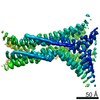
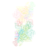
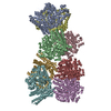
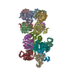
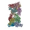
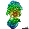
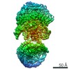
| Title | PARTIAL STRUCTURE OF 16S RNA OF THE SMALL RIBOSOMAL SUBUNIT FROM THERMUS THERMOPHILUS | ||||||||||||
 Components Components |
| ||||||||||||
 Keywords Keywords | RIBOSOME / RIBOSOMES / 30S / THERMUS THERMOPHILUS / 16S RNA | ||||||||||||
| Function / homology |  Function and homology information Function and homology informationribosomal small subunit assembly / small ribosomal subunit / cytosolic small ribosomal subunit / tRNA binding / rRNA binding / structural constituent of ribosome / translation / response to antibiotic / mRNA binding / cytoplasm Similarity search - Function | ||||||||||||
| Biological species |   Thermus thermophilus (bacteria)Geobacillus stearothermophilus (bacteria) Thermus thermophilus (bacteria)Geobacillus stearothermophilus (bacteria) | ||||||||||||
| Method |  X-RAY DIFFRACTION / SYNCHROTRON / MIRAS / Resolution: 4.5 Å X-RAY DIFFRACTION / SYNCHROTRON / MIRAS / Resolution: 4.5 Å | ||||||||||||
 Authors Authors | Tocilj, A. / Schlunzen, F. / Janell, D. / Gluhmann, M. / Hansen, H. / Harms, J. / Bashan, A. / Bartels, H. / Agmon, I. / Franceschi, F. / Yonath, A. | ||||||||||||
 Citation Citation | Journal: Proc.Natl.Acad.Sci.USA / Year: 1999 Title: The small ribosomal subunit from Thermus thermophilus at 4.5 A resolution: pattern fittings and the identification of a functional site. Authors: Tocilj, A. / Schlunzen, F. / Janell, D. / Gluhmann, M. / Hansen, H.A. / Harms, J. / Bashan, A. / Bartels, H. / Agmon, I. / Franceschi, F. / Yonath, A. #1: Journal: Nature / Year: 1992Title: The structure of ribosomal protein-S5 reveals sites of interaction with 16S ribosomal-RNA Authors: Ramakrishnan, V. / White, S. #2: Journal: Structure / Year: 1997Title: The structure of ribosomal protein S7 at 1.9 angstrom resolution reveals a beta-hairpin motif that binds double-stranded nucleic acids Authors: Wimberly, B.T. / White, S. / Ramakrishnan, V. #3: Journal: Acta Crystallogr. / Year: 1953Title: The structure of the 9(18)-heteropoly anion in potassium 9(18)-tungstophosphate, K6(P2W18O62).14H2O Authors: Dawson, B. | ||||||||||||
| History |
|
- Structure visualization
| Structure viewer | Molecule: MolmilJmol/JSmol |
|---|
- Downloads & links
-Download
| PDBx/mmCIF format | 1dv4.cif.gz | 95 KB | Display | PDBx/mmCIF format |
|---|---|---|---|---|
| PDB format | pdb1dv4.ent.gz | 70.4 KB | Display | PDB format |
| PDBx/mmJSON format | 1dv4.json.gz | Tree view | PDBx/mmJSON format | |
| Others |  Other downloads Other downloads |
-Validation report
| Summary document | 1dv4_validation.pdf.gz | 480.7 KB | Display | wwPDB validaton report |
|---|---|---|---|---|
| Full document | 1dv4_full_validation.pdf.gz | 488.6 KB | Display | |
| Data in XML | 1dv4_validation.xml.gz | 4.8 KB | Display | |
| Data in CIF | 1dv4_validation.cif.gz | 10.6 KB | Display | |
| Arichive directory | https://data.pdbj.org/pub/pdb/validation_reports/dv/1dv4ftp://data.pdbj.org/pub/pdb/validation_reports/dv/1dv4 | HTTPS FTP |
-Related structure data
| Similar structure data |
|---|
-Links
 PDBj
PDBj


- Assembly
| Deposited unit | 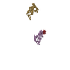
| ||||||||||
|---|---|---|---|---|---|---|---|---|---|---|---|
| 1 |
| ||||||||||
| Unit cell |
|
-Components
| #1: RNA chain | Mass: 70941.203 Da / Num. of mol.: 1 / Source method: isolated from a natural source / Source: (natural) Thermus thermophilus (bacteria) |
|---|---|
| #2: Protein | Mass: 15240.724 Da / Num. of mol.: 1 / Fragment: RESIDUES 4-148 / Source method: isolated from a natural source / Source: (natural) Geobacillus stearothermophilus (bacteria) / References: UniProt: P02357 |
| #3: Protein | Mass: 15342.846 Da / Num. of mol.: 1 / Fragment: RESIDUES 12-146 / Source method: isolated from a natural source / Source: (natural) Thermus thermophilus (bacteria) / References: UniProt: P17291 |
| #4: Chemical | ChemComp-WO2 /   Mass: 4363.030 Da / Num. of mol.: 1 / Source method: obtained synthetically / Formula: O62P2W18 Mass: 4363.030 Da / Num. of mol.: 1 / Source method: obtained synthetically / Formula: O62P2W18 |
-Experimental details
-Experiment
| Experiment | Method: X-RAY DIFFRACTION / Number of used crystals: 1 |
|---|
- Sample preparation
| Crystal | Description: diffraction data were collected during years 1987-2000. |
|---|---|
| Crystal grow | *PLUS Method: other / Details: Yonath, A., (1988) J. Mol. Biol., 203, 831. |
-Data collection
| Diffraction |
| ||||||||||||||||||||||||||||||
|---|---|---|---|---|---|---|---|---|---|---|---|---|---|---|---|---|---|---|---|---|---|---|---|---|---|---|---|---|---|---|---|
| Diffraction source |
| ||||||||||||||||||||||||||||||
| Radiation | Protocol: MAD / Monochromatic (M) / Laue (L): M / Scattering type: x-ray | ||||||||||||||||||||||||||||||
| Radiation wavelength |
| ||||||||||||||||||||||||||||||
| Reflection | Resolution: 4.5→257 Å / Num. all: 85991 / % possible obs: 91.1 % / Observed criterion σ(I): -2.5 / Redundancy: 4 % / Rsym value: 12.5 | ||||||||||||||||||||||||||||||
| Reflection shell | Highest resolution: 4.5 Å / Redundancy: 2 % / % possible all: 91.1 | ||||||||||||||||||||||||||||||
| Reflection | *PLUS Num. obs: 85991 / % possible obs: 93.2 % / Rmerge(I) obs: 0.108 | ||||||||||||||||||||||||||||||
| Reflection shell | *PLUS % possible obs: 91.1 % |


- Processing
| Software |
| ||||||||||||
|---|---|---|---|---|---|---|---|---|---|---|---|---|---|
| Refinement | Method to determine structure: MIRAS / Resolution: 4.5→30 Å / Num. reflection all: 85991 / Num. reflection obs: 85991 / σ(I): 0 Details: Structure was traced manually and no refinement was carried out. Positioning of ribosomal proteins is tentative. The crystallographically determined structure of an 18 W atom cluster (ref. 3) ...Details: Structure was traced manually and no refinement was carried out. Positioning of ribosomal proteins is tentative. The crystallographically determined structure of an 18 W atom cluster (ref. 3) that belongs to the Wative particle was placed around its center of mass in an arbitrary orientation in order to indicate its volume. | ||||||||||||
| Refinement step | Cycle: LAST / Resolution: 4.5→30 Å
| ||||||||||||
| Refinement | *PLUS | ||||||||||||
| Solvent computation | *PLUS | ||||||||||||
| Displacement parameters | *PLUS |
