 Movie
Movie Controller

[English] 日本語
 Yorodumi
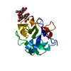
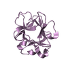
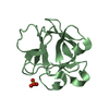
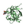
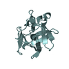
- PDB-1dqo: Crystal structure of the cysteine rich domain of mannose receptor... -
+ Open data

- Basic information
| Entry | Database: PDB / ID: 1dqo | |||||||||
|---|---|---|---|---|---|---|---|---|---|---|
| Title | Crystal structure of the cysteine rich domain of mannose receptor complexed with Acetylgalactosamine-4-sulfate | |||||||||
 Components Components | MANNOSE RECEPTOR | |||||||||
 Keywords Keywords | SUGAR BINDING PROTEIN / beta trefoil / multilectin receptor / pituitary hormones / sulfated carbohydrate | |||||||||
| Function / homology |  Function and homology information Function and homology informationCross-presentation of soluble exogenous antigens (endosomes) / cargo receptor activity / mannose binding / receptor-mediated endocytosis / cellular response to interleukin-4 / cellular response to type II interferon / transmembrane signaling receptor activity / signaling receptor activity / cellular response to lipopolysaccharide / endosome membrane ...Cross-presentation of soluble exogenous antigens (endosomes) / cargo receptor activity / mannose binding / receptor-mediated endocytosis / cellular response to interleukin-4 / cellular response to type II interferon / transmembrane signaling receptor activity / signaling receptor activity / cellular response to lipopolysaccharide / endosome membrane / cell surface / plasma membraneSimilarity search - Function | |||||||||
| Biological species |  Mus musculus (house mouse) Mus musculus (house mouse) | |||||||||
| Method | X-RAY DIFFRACTION / MOLECULAR REPLACEMENT / Resolution: 2.2 Å | |||||||||
 Authors Authors | Liu, Y. / Chirino, A.J. / Misulovin, Z. / Leteux, C. / Feizi, T. / Nussenzweig, M.C. / Bjorkman, P.J. | |||||||||
 Citation Citation | Journal: J.Exp.Med. / Year: 2000 Title: Crystal structure of the cysteine-rich domain of mannose receptor complexed with a sulfated carbohydrate ligand. Authors: Liu, Y. / Chirino, A.J. / Misulovin, Z. / Leteux, C. / Feizi, T. / Nussenzweig, M.C. / Bjorkman, P.J. | |||||||||
| History |
|
- Structure visualization
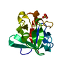
| Structure viewer | Molecule: MolmilJmol/JSmol |
|---|
- Downloads & links
-Download
| PDBx/mmCIF format | 1dqo.cif.gz | 44.7 KB | Display | PDBx/mmCIF format |
|---|---|---|---|---|
| PDB format | pdb1dqo.ent.gz | 30.1 KB | Display | PDB format |
| PDBx/mmJSON format | 1dqo.json.gz | Tree view | PDBx/mmJSON format | |
| Others |  Other downloads Other downloads |
-Validation report
| Arichive directory | https://data.pdbj.org/pub/pdb/validation_reports/dq/1dqoftp://data.pdbj.org/pub/pdb/validation_reports/dq/1dqo | HTTPS FTP |
|---|
-Related structure data
| Related structure data | 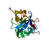 1dqgSC S: Starting model for refinement C: citing same article ( |
|---|---|
| Similar structure data |


-Links
 PDBj
PDBj


- Assembly
| Deposited unit | 
| ||||||||
|---|---|---|---|---|---|---|---|---|---|
| 1 |
| ||||||||
| Unit cell |
|
-Components
| #1: Protein | Mass: 15558.486 Da / Num. of mol.: 1 / Fragment: CYSTEINE RICH DOMAIN Source method: isolated from a genetically manipulated source Source: (gene. exp.) Mus musculus (house mouse) / Cell: MACROPHAGE / Cell line: EPITHELIAL CELL / Production host:  Homo sapiens (human) / References: UniProt: Q61830 Homo sapiens (human) / References: UniProt: Q61830 |
|---|---|
| #2: Sugar | ChemComp-ASG / 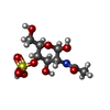  Type: D-saccharide, beta linking / Mass: 301.271 Da / Num. of mol.: 1 Type: D-saccharide, beta linking / Mass: 301.271 Da / Num. of mol.: 1Source method: isolated from a genetically manipulated source Formula: C8H15NO9S |
| #3: Water | ChemComp-HOH / Water Mass: 18.015 Da / Num. of mol.: 153 / Source method: isolated from a natural source / Formula: H2O Mass: 18.015 Da / Num. of mol.: 153 / Source method: isolated from a natural source / Formula: H2O |
-Experimental details
-Experiment
| Experiment | Method: X-RAY DIFFRACTION / Number of used crystals: 1 |
|---|
- Sample preparation
| Crystal | Density Matthews: 2.64 Å3/Da / Density % sol: 53.33 % | ||||||||||||||||||||||||||||||||||||
|---|---|---|---|---|---|---|---|---|---|---|---|---|---|---|---|---|---|---|---|---|---|---|---|---|---|---|---|---|---|---|---|---|---|---|---|---|---|
| Crystal grow | Temperature: 277 K / Method: vapor diffusion, hanging drop / pH: 6.5 Details: PEG8000, ammonium sulfate, pH 6.5, VAPOR DIFFUSION, HANGING DROP, temperature 277K | ||||||||||||||||||||||||||||||||||||
| Crystal grow | *PLUS Temperature: 4 ℃ / pH: 7.4 | ||||||||||||||||||||||||||||||||||||
| Components of the solutions | *PLUS
|
-Data collection
| Diffraction | Mean temperature: 120 K |
|---|---|
| Diffraction source | Source: ROTATING ANODE / Type: RIGAKU RU200 / Wavelength: 1.5418 |
| Detector | Type: RIGAKU RAXIS II / Detector: IMAGE PLATE / Date: Oct 30, 1998 |
| Radiation | Protocol: SINGLE WAVELENGTH / Monochromatic (M) / Laue (L): M / Scattering type: x-ray |
| Radiation wavelength | Wavelength: 1.5418 Å / Relative weight: 1 |
| Reflection | Resolution: 2.2→25 Å / Num. all: 8984 / Num. obs: 8984 / % possible obs: 97.5 % / Observed criterion σ(F): 0 / Observed criterion σ(I): 0 / Redundancy: 9.6 % / Biso Wilson estimate: 24.1 Å2 / Rmerge(I) obs: 0.045 / Net I/σ(I): 24.6 |
| Reflection shell | Resolution: 2.2→2.28 Å / Redundancy: 3.8 % / Rmerge(I) obs: 0.105 / % possible all: 95.5 |
| Reflection | *PLUS Num. measured all: 86630 |
| Reflection shell | *PLUS % possible obs: 95.5 % / Mean I/σ(I) obs: 11.8 |
- Processing
| Software |
| ||||||||||||||||||||
|---|---|---|---|---|---|---|---|---|---|---|---|---|---|---|---|---|---|---|---|---|---|
| Refinement | Method to determine structure: MOLECULAR REPLACEMENT Starting model: 1DQG Resolution: 2.2→25 Å / σ(F): 0 / σ(I): 0 / Stereochemistry target values: Engh & Huber
| ||||||||||||||||||||
| Refinement step | Cycle: LAST / Resolution: 2.2→25 Å
| ||||||||||||||||||||
| Refine LS restraints |
| ||||||||||||||||||||
| Software | *PLUS Name: CNS / Classification: refinement | ||||||||||||||||||||
| Refinement | *PLUS Highest resolution: 2.2 Å / Lowest resolution: 25 Å / σ(F): 0 | ||||||||||||||||||||
| Solvent computation | *PLUS | ||||||||||||||||||||
| Displacement parameters | *PLUS |
