 Movie
Movie Controller

[English] 日本語
 Yorodumi
- PDB-1db1: CRYSTAL STRUCTURE OF THE NUCLEAR RECEPTOR FOR VITAMIN D COMPLEXED... -
+ Open data

- Basic information
| Entry | Database: PDB / ID: 1db1 | ||||||
|---|---|---|---|---|---|---|---|
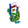
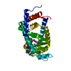
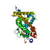
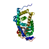
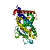
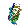
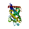
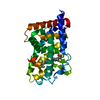
| Title | CRYSTAL STRUCTURE OF THE NUCLEAR RECEPTOR FOR VITAMIN D COMPLEXED TO VITAMIN D | ||||||
 Components Components | VITAMIN D NUCLEAR RECEPTOR | ||||||
 Keywords Keywords |  GENE REGULATION / COMPLEX GENE REGULATION / COMPLEX | ||||||
| Function / homology |  Function and homology informationregulation of calcidiol 1-monooxygenase activity / positive regulation of vitamin D 24-hydroxylase activity / bile acid nuclear receptor activity / response to bile acid / Vitamin D (calciferol) metabolism / phosphate ion transmembrane transport / apoptotic process involved in mammary gland involution / positive regulation of apoptotic process involved in mammary gland involution / calcitriol binding / lithocholic acid binding ...regulation of calcidiol 1-monooxygenase activity / positive regulation of vitamin D 24-hydroxylase activity / bile acid nuclear receptor activity / response to bile acid / Vitamin D (calciferol) metabolism / phosphate ion transmembrane transport / apoptotic process involved in mammary gland involution / positive regulation of apoptotic process involved in mammary gland involution / calcitriol binding / lithocholic acid binding / positive regulation of keratinocyte differentiation / positive regulation of vitamin D receptor signaling pathway / vitamin D receptor signaling pathway / bile acid signaling pathway / intestinal absorption / mammary gland branching involved in pregnancy / decidualization / negative regulation of keratinocyte proliferation / positive regulation of bone mineralization / nuclear retinoid X receptor binding / lactation / skeletal system development / SUMOylation of intracellular receptors / mRNA transcription by RNA polymerase II / cell morphogenesis / intracellular calcium ion homeostasis / Nuclear Receptor transcription pathway / RNA polymerase II transcription regulator complex / nuclear receptor activity / calcium ion transport / cell differentiation / receptor complex / DNA-binding transcription factor activity, RNA polymerase II-specific / RNA polymerase II cis-regulatory region sequence-specific DNA binding / negative regulation of cell population proliferation / negative regulation of DNA-templated transcription / chromatin / positive regulation of gene expression / negative regulation of transcription by RNA polymerase II / positive regulation of transcription by RNA polymerase II / DNA binding / zinc ion binding / nucleoplasm / nucleus / cytosol Function and homology informationregulation of calcidiol 1-monooxygenase activity / positive regulation of vitamin D 24-hydroxylase activity / bile acid nuclear receptor activity / response to bile acid / Vitamin D (calciferol) metabolism / phosphate ion transmembrane transport / apoptotic process involved in mammary gland involution / positive regulation of apoptotic process involved in mammary gland involution / calcitriol binding / lithocholic acid binding ...regulation of calcidiol 1-monooxygenase activity / positive regulation of vitamin D 24-hydroxylase activity / bile acid nuclear receptor activity / response to bile acid / Vitamin D (calciferol) metabolism / phosphate ion transmembrane transport / apoptotic process involved in mammary gland involution / positive regulation of apoptotic process involved in mammary gland involution / calcitriol binding / lithocholic acid binding / positive regulation of keratinocyte differentiation / positive regulation of vitamin D receptor signaling pathway / vitamin D receptor signaling pathway / bile acid signaling pathway / intestinal absorption / mammary gland branching involved in pregnancy / decidualization / negative regulation of keratinocyte proliferation / positive regulation of bone mineralization / nuclear retinoid X receptor binding / lactation / skeletal system development / SUMOylation of intracellular receptors / mRNA transcription by RNA polymerase II / cell morphogenesis / intracellular calcium ion homeostasis / Nuclear Receptor transcription pathway / RNA polymerase II transcription regulator complex / nuclear receptor activity / calcium ion transport / cell differentiation / receptor complex / DNA-binding transcription factor activity, RNA polymerase II-specific / RNA polymerase II cis-regulatory region sequence-specific DNA binding / negative regulation of cell population proliferation / negative regulation of DNA-templated transcription / chromatin / positive regulation of gene expression / negative regulation of transcription by RNA polymerase II / positive regulation of transcription by RNA polymerase II / DNA binding / zinc ion binding / nucleoplasm / nucleus / cytosolSimilarity search - Function | ||||||
| Biological species |  Homo sapiens (human) Homo sapiens (human) | ||||||
| Method | X-RAY DIFFRACTION / SYNCHROTRON / Resolution: 1.8 Å | ||||||
 Authors Authors | Rochel, N. / Wurtz, J.M. / Mitschler, A. / Klaholz, B. / Moras, D. | ||||||
 Citation Citation | Journal: Mol.Cell / Year: 2000 Title: The crystal structure of the nuclear receptor for vitamin D bound to its natural ligand. Authors: Rochel, N. / Wurtz, J.M. / Mitschler, A. / Klaholz, B. / Moras, D. | ||||||
| History |
|
- Structure visualization
| Structure viewer | Molecule: MolmilJmol/JSmol |
|---|
- Downloads & links
-Download
| PDBx/mmCIF format | 1db1.cif.gz | 66.1 KB | Display | PDBx/mmCIF format |
|---|---|---|---|---|
| PDB format | pdb1db1.ent.gz | 48.3 KB | Display | PDB format |
| PDBx/mmJSON format | 1db1.json.gz | Tree view | PDBx/mmJSON format | |
| Others |  Other downloads Other downloads |
-Validation report
| Arichive directory | https://data.pdbj.org/pub/pdb/validation_reports/db/1db1ftp://data.pdbj.org/pub/pdb/validation_reports/db/1db1 | HTTPS FTP |
|---|
-Related structure data
| Similar structure data |
|---|


-Links
 PDBj
PDBj

- Assembly
| Deposited unit | 
| ||||||||
|---|---|---|---|---|---|---|---|---|---|
| 1 |
| ||||||||
| Unit cell |
|
-Components
| #1: Protein | Mass: 29391.871 Da / Num. of mol.: 1 / Fragment: LIGAND BINDING DOMAIN Source method: isolated from a genetically manipulated source Source: (gene. exp.) Homo sapiens (human) / Plasmid: PET15B / Production host:  Escherichia coli (E. coli) / References: UniProt: P11473 Escherichia coli (E. coli) / References: UniProt: P11473 |
|---|---|
| #2: Chemical | ChemComp-VDX / Calcitriol  Mass: 416.636 Da / Num. of mol.: 1 / Source method: obtained synthetically / Formula: C27H44O3 Mass: 416.636 Da / Num. of mol.: 1 / Source method: obtained synthetically / Formula: C27H44O3 |
| #3: Water | ChemComp-HOH / Water Mass: 18.015 Da / Num. of mol.: 164 / Source method: isolated from a natural source / Formula: H2O Mass: 18.015 Da / Num. of mol.: 164 / Source method: isolated from a natural source / Formula: H2O |
-Experimental details
-Experiment
| Experiment | Method: X-RAY DIFFRACTION / Number of used crystals: 4 |
|---|
- Sample preparation
| Crystal | Density Matthews: 2.69 Å3/Da / Density % sol: 54.2 % | ||||||||||||||||||||||||||||||||||||||||
|---|---|---|---|---|---|---|---|---|---|---|---|---|---|---|---|---|---|---|---|---|---|---|---|---|---|---|---|---|---|---|---|---|---|---|---|---|---|---|---|---|---|
| Crystal grow | Temperature: 298 K / Method: vapor diffusion, hanging drop / pH: 6 Details: ammonium sulfate, mes, pH 6.0, VAPOR DIFFUSION, HANGING DROP, temperature 298K | ||||||||||||||||||||||||||||||||||||||||
| Crystal grow | *PLUS Temperature: 4 ℃ | ||||||||||||||||||||||||||||||||||||||||
| Components of the solutions | *PLUS
|
-Data collection
| Diffraction | Mean temperature: 298 K |
|---|---|
| Diffraction source | Source: SYNCHROTRON / Site: EMBL/DESY, HAMBURG  / Beamline: BW7B / Wavelength: 0.8345 / Beamline: BW7B / Wavelength: 0.8345 |
| Detector | Type: MARRESEARCH / Detector: IMAGE PLATE / Date: Jul 11, 1998 |
| Radiation | Protocol: SINGLE WAVELENGTH / Monochromatic (M) / Laue (L): M / Scattering type: x-ray |
| Radiation wavelength | Wavelength: 0.8345 Å / Relative weight: 1 |
| Reflection | Resolution: 1.8→20 Å / Num. all: 30148 / Num. obs: 29434 / % possible obs: 97.4 % / Observed criterion σ(F): 1 / Observed criterion σ(I): 1 / Redundancy: 4.1 % / Biso Wilson estimate: 26 Å2 / Rmerge(I) obs: 0.061 / Net I/σ(I): 22.1 |
| Reflection shell | Resolution: 1.8→1.84 Å / Redundancy: 2.8 % / Rmerge(I) obs: 0.24 / % possible all: 95.3 |
| Reflection shell | *PLUS % possible obs: 95.3 % |
- Processing
| Software |
| ||||||||||||||||||||||||||||||
|---|---|---|---|---|---|---|---|---|---|---|---|---|---|---|---|---|---|---|---|---|---|---|---|---|---|---|---|---|---|---|---|
| Refinement | Resolution: 1.8→20 Å / σ(F): 0 / σ(I): 0 / Stereochemistry target values: Engh & Huber
| ||||||||||||||||||||||||||||||
| Refinement step | Cycle: LAST / Resolution: 1.8→20 Å
| ||||||||||||||||||||||||||||||
| Software | *PLUS Name: 'CNS' / Classification: refinement | ||||||||||||||||||||||||||||||
| Refine LS restraints | *PLUS
|
