[English] 日本語
Yorodumi
- PDB-1rl6: RIBOSOMAL PROTEIN L6 -

+
Open data


ID or keywords:

Loading...

-
Basic information

Entry
Database: PDB / ID: 1rl6
TitleRIBOSOMAL PROTEIN L6
ComponentsPROTEIN (RIBOSOMAL PROTEIN L6)
KeywordsRNA BINDING PROTEIN / RNA-BINDING PROTEIN / GENTAMICIN RESISTANCE / RIBOSOMAL PROTEIN / ALPHA/BETA PROTEIN
Function / homology
Function and homology information


rRNA binding / ribosome / structural constituent of ribosome / translation / ribonucleoprotein complex
Similarity search - Function
Ribosomal protein L6 / Outer Surface Protein A; domain 3 / Ribosomal protein L6, conserved site / Ribosomal protein L6 signature 1. / Ribosomal protein L6, bacterial-type / Ribosomal protein L6, alpha-beta domain / Ribosomal protein L6 / Ribosomal protein L6, alpha-beta domain superfamily / Ribosomal protein L6 / Alpha-Beta Complex / Alpha Beta
Similarity search - Domain/homology
Large ribosomal subunit protein uL6
Similarity search - Component
Biological speciesGeobacillus stearothermophilus (bacteria)
MethodX-RAY DIFFRACTION / MIR / Resolution: 2 Å
AuthorsGolden, B.L. / Davies, C. / Ramakrishnan, V. / White, S.W.
Citation
Journal: EMBO J. / Year: 1993
Title: Ribosomal protein L6: structural evidence of gene duplication from a primitive RNA binding protein.
Authors: Golden, B.L. / Ramakrishnan, V. / White, S.W.
#1: Journal: J.Mol.Biol. / Year: 1998
Title: Ribosomal proteins S5 and L6: high-resolution crystal structures and roles in protein synthesis and antibiotic resistance.
Authors: Davies, C. / Bussiere, D.E. / Golden, B.L. / Porter, S.J. / Ramakrishnan, V. / White, S.W.
History
DepositionJan 14, 1999Processing site: RCSB
Revision 1.0Feb 2, 1999Provider: repository / Type: Initial release
Revision 1.1Apr 26, 2008Group: Version format compliance
Revision 1.2Jul 13, 2011Group: Version format compliance
Revision 1.3Oct 30, 2019Group: Data collection / Database references / Structure summary
Category: citation / struct_keywords
Item: _citation.page_last / _citation.pdbx_database_id_PubMed ..._citation.page_last / _citation.pdbx_database_id_PubMed / _citation.title / _struct_keywords.text
Revision 1.4Dec 27, 2023Group: Data collection / Database references / Category: chem_comp_atom / chem_comp_bond / database_2
Item: _database_2.pdbx_DOI / _database_2.pdbx_database_accession

-
Structure visualization

Structure viewerMolecule:
MolmilJmol/JSmol

Downloads & links

-
Assembly

Deposited unit
A: PROTEIN (RIBOSOMAL PROTEIN L6)


Theoretical massNumber of molelcules
Total (without water)19,2021
Polymers19,2021
Non-polymers00
Water2,522140
1


  • Idetical with deposited unit
  • defined by author
TypeNameSymmetry operationNumber
identity operation1_555x,y,z1
Unit cell
Length a, b, c (Å)71.800, 71.800, 124.500
Angle α, β, γ (deg.)90.00, 90.00, 120.00
Int Tables number178
Cell settinghexagonal
Space group name H-MP6122

-
Components

#1: Protein PROTEIN (RIBOSOMAL PROTEIN L6)


Mass: 19202.123 Da / Num. of mol.: 1
Source method: isolated from a genetically manipulated source
Details: LOCATED IN THE LARGE 50S RIBOSOMAL SUBUNIT BELOW THE L7/L12 STALK AND CLOSE TO THE GTPASE CENTER
Source: (gene. exp.) Geobacillus stearothermophilus (bacteria)
Cellular location: RIBOSOME / Plasmid: PET-13A / Species (production host): Escherichia coli / Production host: Escherichia coli BL21(DE3) (bacteria) / Strain (production host): BL21(DE3) / References: UniProt: P02391
#2: Water ChemComp-HOH / water / Water


Mass: 18.015 Da / Num. of mol.: 140 / Source method: isolated from a natural source / Formula: H2O
Compound details
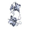
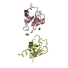
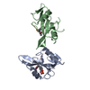
THE PROTEIN CONTAINS TWO DOMAINS OF APPROXIMATELY EQUAL SIZE. THE DOMAINS HAVE VERY SIMILAR ALPHA ...THE PROTEIN CONTAINS TWO DOMAINS OF APPROXIMATELY EQUAL SIZE. THE DOMAINS HAVE VERY SIMILAR ALPHA BETA STRUCTURES AND SECONDARY STRUCTURE TOPOLOGIES AND APPEAR TO HAVE ARISEN BY GENE DUPLICATION. EACH DOMAIN IS ALSO HOMOLOGOUS TO THE RNA RECOGNITION MOTIF (RRM), WITH THE C-TERMINAL DOMAIN BEING THE MOST SIMILAR.

-
Experimental details

-
Experiment

ExperimentMethod: X-RAY DIFFRACTION / Number of used crystals: 1

-
Sample preparation

CrystalDensity Matthews: 2.4 Å3/Da / Density % sol: 50 %
Description: MUTANT V124C WAS CONSTRUCTED FOR THE MERCURY DERIVATIVE
Crystal growpH: 7.6 / Details: pH 7.6
Components of the solutions
IDNameCrystal-IDSol-ID
1NA/K PHOSPHATE11
2BETA MERCAPTOETHANOL11
31,4-DIOXANE11
Crystal grow
*PLUS
Method: vapor diffusion, hanging drop / PH range low: 7.8 / PH range high: 7.2
Components of the solutions
*PLUS
IDConc.Common nameCrystal-IDSol-ID
110-15 mg/mlprotein1drop
20.6 Msodium potassium phosphate1drop
33 mMbeta-mercaptoethanol1drop
43 %(v/v)1,4-dioxane1drop
51.6-1.8 Msodium potassium phospahte1reservoir
63 mMbeta-mercaptoethanol1reservoir
73 %1,4-dioxane1reservoir

-
Data collection

DiffractionMean temperature: 293 K
Diffraction sourceSource: ROTATING ANODE / Type: RIGAKU RU300 / Wavelength: 1.5418
DetectorType: RIGAKU / Detector: IMAGE PLATE / Details: COLLIMATOR
RadiationMonochromator: GRAPHITE / Protocol: SINGLE WAVELENGTH / Monochromatic (M) / Laue (L): M / Scattering type: x-ray
Radiation wavelengthWavelength: 1.5418 Å / Relative weight: 1
ReflectionResolution: 2→20 Å / Num. obs: 13166 / % possible obs: 97.5 % / Redundancy: 3 % / Biso Wilson estimate: 19.7 Å2 / Rmerge(I) obs: 0.08
Reflection shellResolution: 2→2.09 Å / % possible all: 97.4

-
Processing

Software
NameVersionClassification
PHASESphasing
X-PLOR3.851refinement
DENZOdata reduction
SCALEPACKdata scaling
RefinementMethod to determine structure: MIR / Resolution: 2→20 Å / Rfactor Rfree error: 0.008 / Data cutoff high absF: 18251945.8 / Data cutoff low absF: 0 / Isotropic thermal model: RESTRAINED / Cross valid method: THROUGHOUT / σ(F): 0
RfactorNum. reflection% reflectionSelection details
Rfree0.302 1288 9.8 %RANDOM
Rwork0.257 ---
all-13142 --
obs-13142 97.8 %-
Displacement parametersBiso mean: 38.7 Å2
Baniso -1Baniso -2Baniso -3
1--0.07 Å2-1.85 Å20 Å2
2---0.07 Å20 Å2
3---0.13 Å2
Refine analyze
FreeObs
Luzzati coordinate error0.4 Å0.33 Å
Luzzati d res low-5 Å
Luzzati sigma a0.36 Å0.36 Å
Refinement stepCycle: LAST / Resolution: 2→20 Å
ProteinNucleic acidLigandSolventTotal
Num. atoms1251 0 0 140 1391
Refine LS restraints
Refine-IDTypeDev idealDev ideal target
X-RAY DIFFRACTIONx_bond_d0.014
X-RAY DIFFRACTIONx_bond_d_na
X-RAY DIFFRACTIONx_bond_d_prot
X-RAY DIFFRACTIONx_angle_d
X-RAY DIFFRACTIONx_angle_d_na
X-RAY DIFFRACTIONx_angle_d_prot
X-RAY DIFFRACTIONx_angle_deg1.6
X-RAY DIFFRACTIONx_angle_deg_na
X-RAY DIFFRACTIONx_angle_deg_prot
X-RAY DIFFRACTIONx_dihedral_angle_d26.5
X-RAY DIFFRACTIONx_dihedral_angle_d_na
X-RAY DIFFRACTIONx_dihedral_angle_d_prot
X-RAY DIFFRACTIONx_improper_angle_d1.43
X-RAY DIFFRACTIONx_improper_angle_d_na
X-RAY DIFFRACTIONx_improper_angle_d_prot
X-RAY DIFFRACTIONx_mcbond_it2.041.5
X-RAY DIFFRACTIONx_mcangle_it3.282
X-RAY DIFFRACTIONx_scbond_it5.022
X-RAY DIFFRACTIONx_scangle_it7.122.5
LS refinement shellResolution: 2→2.13 Å / Rfactor Rfree error: 0.03 / Total num. of bins used: 6
RfactorNum. reflection% reflection
Rfree0.41 186 8.9 %
Rwork0.345 1893 -
obs--96.2 %
Xplor fileSerial no: 1 / Param file: PARAM19X.PRO / Topol file: TOPH19X.PRO
Software
*PLUS
Name: X-PLOR / Version: 3.851 / Classification: refinement
Refinement
*PLUS
Highest resolution: 2 Å / Lowest resolution: 20 Å / σ(F): 0 / % reflection Rfree: 9.8 % / Rfactor obs: 0.257
Solvent computation
*PLUS
Displacement parameters
*PLUS
Biso mean: 38.7 Å2
Refine LS restraints
*PLUS
Refine-IDTypeDev idealDev ideal target
X-RAY DIFFRACTIONx_angle_deg1.6
X-RAY DIFFRACTIONx_dihedral_angle_d
X-RAY DIFFRACTIONx_dihedral_angle_deg26.5
X-RAY DIFFRACTIONx_improper_angle_d
X-RAY DIFFRACTIONx_improper_angle_deg1.43
X-RAY DIFFRACTIONx_mcbond_it1.5
X-RAY DIFFRACTIONx_scbond_it2
X-RAY DIFFRACTIONx_mcangle_it2
X-RAY DIFFRACTIONx_scangle_it2.5
LS refinement shell
*PLUS
Rfactor Rfree: 0.41 / % reflection Rfree: 8.9 % / Rfactor Rwork: 0.345

+
About Yorodumi

-
News

-
Feb 9, 2022. New format data for meta-information of EMDB entries

New format data for meta-information of EMDB entries

  • Version 3 of the EMDB header file is now the official format.
  • The previous official version 1.9 will be removed from the archive.

Related info.:EMDB header

External links:wwPDB to switch to version 3 of the EMDB data model

-
Aug 12, 2020. Covid-19 info

Covid-19 info

URL: https://pdbj.org/emnavi/covid19.php

New page: Covid-19 featured information page in EM Navigator.

Related info.:Covid-19 info / Mar 5, 2020. Novel coronavirus structure data

+
Mar 5, 2020. Novel coronavirus structure data

Novel coronavirus structure data

Related info.:Yorodumi Speices / Aug 12, 2020. Covid-19 info

External links:COVID-19 featured content - PDBj / Molecule of the Month (242):Coronavirus Proteases

+
Jan 31, 2019. EMDB accession codes are about to change! (news from PDBe EMDB page)

EMDB accession codes are about to change! (news from PDBe EMDB page)

  • The allocation of 4 digits for EMDB accession codes will soon come to an end. Whilst these codes will remain in use, new EMDB accession codes will include an additional digit and will expand incrementally as the available range of codes is exhausted. The current 4-digit format prefixed with “EMD-” (i.e. EMD-XXXX) will advance to a 5-digit format (i.e. EMD-XXXXX), and so on. It is currently estimated that the 4-digit codes will be depleted around Spring 2019, at which point the 5-digit format will come into force.
  • The EM Navigator/Yorodumi systems omit the EMD- prefix.

Related info.:Q: What is EMD? / ID/Accession-code notation in Yorodumi/EM Navigator

External links:EMDB Accession Codes are Changing Soon! / Contact to PDBj

+
Jul 12, 2017. Major update of PDB

Major update of PDB

  • wwPDB released updated PDB data conforming to the new PDBx/mmCIF dictionary.
  • This is a major update changing the version number from 4 to 5, and with Remediation, in which all the entries are updated.
  • In this update, many items about electron microscopy experimental information are reorganized (e.g. em_software).
  • Now, EM Navigator and Yorodumi are based on the updated data.

External links:wwPDB Remediation / Enriched Model Files Conforming to OneDep Data Standards Now Available in the PDB FTP Archive

-
Yorodumi

Thousand views of thousand structures

  • Yorodumi is a browser for structure data from EMDB, PDB, SASBDB, etc.
  • This page is also the successor to EM Navigator detail page, and also detail information page/front-end page for Omokage search.
  • The word "yorodu" (or yorozu) is an old Japanese word meaning "ten thousand". "mi" (miru) is to see.

Related info.:EMDB / PDB / SASBDB / Comparison of 3 databanks / Yorodumi Search / Aug 31, 2016. New EM Navigator & Yorodumi / Yorodumi Papers / Jmol/JSmol / Function and homology information / Changes in new EM Navigator and Yorodumi

Read more