 Movie
Movie Controller

[English] 日本語
 Yorodumi
- PDB-1pkp: THE STRUCTURE OF RIBOSOMAL PROTEIN S5 REVEALS SITES OF INTERACTIO... -
+ Open data

- Basic information
| Entry | Database: PDB / ID: 1pkp | ||||||
|---|---|---|---|---|---|---|---|
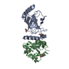
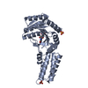
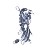
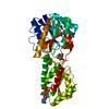
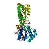
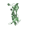
| Title | THE STRUCTURE OF RIBOSOMAL PROTEIN S5 REVEALS SITES OF INTERACTION WITH 16S RRNA | ||||||
 Components Components | RIBOSOMAL PROTEIN S5 | ||||||
 Keywords Keywords | RIBOSOMAL PROTEIN | ||||||
| Function / homology |  Function and homology information Function and homology informationsmall ribosomal subunit / rRNA binding / structural constituent of ribosome / translation / response to antibiotic / cytoplasmSimilarity search - Function | ||||||
| Biological species |   Geobacillus stearothermophilus (bacteria) Geobacillus stearothermophilus (bacteria) | ||||||
| Method | X-RAY DIFFRACTION / Resolution: 2.8 Å | ||||||
 Authors Authors | Ramakrishnan, V. / White, S.W. | ||||||
 Citation Citation | Journal: Nature / Year: 1992 Title: The structure of ribosomal protein S5 reveals sites of interaction with 16S rRNA. Authors: Ramakrishnan, V. / White, S.W. #1: Journal: J.Mol.Biol. / Year: 1988Title: Model for the Three-Dimensional Folding of 16S Ribosomal RNA Authors: Stern, S. / Weiser, B. / Noller, H.F. #2: Journal: FEBS Lett. / Year: 1983Title: Proteins of the Bacillus Stearothermophilus Ribosome: A 5A Structure Analysis of Protein S5 Authors: White, S.W. / Appelt, K. / Dijk, J. / Wilson, K.S. #3: Journal: J.Biol.Chem. / Year: 1983Title: Proteins of the Bacillus Stearothermophilus Ribosome. Crystallization of Proteins L30 and S5 Authors: Appelt, K. / White, S.W. / Wilson, K.S. #4: Journal: Mol.Gen.Genet. / Year: 1975Title: Effect of Different Mutations in Ribosomal Protein S5 of Escherichia Coli on Translational Fidelity Authors: Piepersberg, W. / Bock, A. / Wittmann, H.-G. #5: Journal: Mol.Gen.Genet. / Year: 1975Title: Genetic Position and Amino Acid Replacements of Several Mutations in Ribosomal Protein S5 from Escherichia Coli Authors: Piepersberg, W. / Bock, A. / Yaguchi, M. / Wittmann, H.-G. | ||||||
| History |
|
- Structure visualization
| Structure viewer | Molecule: MolmilJmol/JSmol |
|---|
- Downloads & links
-Download
| PDBx/mmCIF format | 1pkp.cif.gz | 36.7 KB | Display | PDBx/mmCIF format |
|---|---|---|---|---|
| PDB format | pdb1pkp.ent.gz | 26 KB | Display | PDB format |
| PDBx/mmJSON format | 1pkp.json.gz | Tree view | PDBx/mmJSON format | |
| Others |  Other downloads Other downloads |
-Validation report
| Arichive directory | https://data.pdbj.org/pub/pdb/validation_reports/pk/1pkpftp://data.pdbj.org/pub/pdb/validation_reports/pk/1pkp | HTTPS FTP |
|---|
-Related structure data
| Similar structure data |
|---|


-Links
 PDBj
PDBj- Assembly
| Deposited unit | 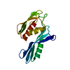
| ||||||||
|---|---|---|---|---|---|---|---|---|---|
| 1 | 
| ||||||||
| Unit cell |
|
-Components
| #1: Protein | Mass: 15914.577 Da / Num. of mol.: 1 Source method: isolated from a genetically manipulated source Source: (gene. exp.) Geobacillus stearothermophilus (bacteria)Gene: BACILLUS STEAROTHERMOPHILUS / Gene (production host): BACILLUS STEAROTHERMOPHILUS / References: UniProt: P02357 |
|---|
-Experimental details
-Experiment
| Experiment | Method: X-RAY DIFFRACTION |
|---|
- Sample preparation
| Crystal | Density Matthews: 3.5 Å3/Da / Density % sol: 64.86 % | ||||||||||||||||||
|---|---|---|---|---|---|---|---|---|---|---|---|---|---|---|---|---|---|---|---|
| Crystal grow | *PLUS Method: unknown / PH range low: 8.2 / PH range high: 7.2 | ||||||||||||||||||
| Components of the solutions | *PLUS
|
-Data collection
| Radiation | Scattering type: x-ray |
|---|---|
| Radiation wavelength | Relative weight: 1 |
| Reflection | *PLUS Highest resolution: 2.7 Å / Num. obs: 5829 / Num. measured all: 14806 / Rmerge(I) obs: 0.051 |
- Processing
| Software |
| ||||||||||||||||||||||||||||||||||||||||||||||||||||||||||||
|---|---|---|---|---|---|---|---|---|---|---|---|---|---|---|---|---|---|---|---|---|---|---|---|---|---|---|---|---|---|---|---|---|---|---|---|---|---|---|---|---|---|---|---|---|---|---|---|---|---|---|---|---|---|---|---|---|---|---|---|---|---|
| Refinement | Resolution: 2.8→6 Å / Rfactor Rwork: 0.22 / Rfactor obs: 0.22 / σ(F): 0 | ||||||||||||||||||||||||||||||||||||||||||||||||||||||||||||
| Refinement step | Cycle: LAST / Resolution: 2.8→6 Å
| ||||||||||||||||||||||||||||||||||||||||||||||||||||||||||||
| Refine LS restraints |
| ||||||||||||||||||||||||||||||||||||||||||||||||||||||||||||
| Software | *PLUS Name: X-PLOR / Classification: refinement | ||||||||||||||||||||||||||||||||||||||||||||||||||||||||||||
| Refinement | *PLUS Highest resolution: 2.8 Å / Lowest resolution: 6 Å / σ(F): 0 / Rfactor all: 0.22 | ||||||||||||||||||||||||||||||||||||||||||||||||||||||||||||
| Solvent computation | *PLUS | ||||||||||||||||||||||||||||||||||||||||||||||||||||||||||||
| Displacement parameters | *PLUS | ||||||||||||||||||||||||||||||||||||||||||||||||||||||||||||
| Refine LS restraints | *PLUS Type: x_angle_d / Dev ideal: 3.74 |
