 Movie
Movie Controller


+ Open data

- Basic information
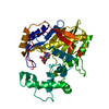
| Entry | Database: PDB / ID: 1mms | ||||||
|---|---|---|---|---|---|---|---|
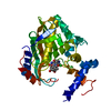
| Title | Crystal structure of the ribosomal PROTEIN L11-RNA complex | ||||||
 Components Components |
| ||||||
 Keywords Keywords |  RIBOSOME / RNA-PROTEIN COMPLEX / RNA / TRANSLOCATION / THIOSTREPTON RIBOSOME / RNA-PROTEIN COMPLEX / RNA / TRANSLOCATION / THIOSTREPTON | ||||||
| Function / homology |  Function and homology information Function and homology informationlarge ribosomal subunit rRNA binding / cytosolic large ribosomal subunit / structural constituent of ribosome / translationSimilarity search - Function | ||||||
| Biological species |   Thermotoga maritima (bacteria) Thermotoga maritima (bacteria) | ||||||
| Method | X-RAY DIFFRACTION / SYNCHROTRON / MAD / Resolution: 2.57 Å | ||||||
 Authors Authors | Wimberly, B.T. / Guymon, R. / Mccutcheon, J.P. / White, S.W. / Ramakrishnan, V. | ||||||
 Citation Citation | Journal: Cell / Year: 1999 Title: A detailed view of a ribosomal active site: the structure of the L11-RNA complex. Authors: B T Wimberly / R Guymon / J P McCutcheon / S W White / V Ramakrishnan /  Abstract: We report the crystal structure of a 58 nucleotide fragment of 23S ribosomal RNA bound to ribosomal protein L11. This highly conserved ribonucleoprotein domain is the target for the thiostrepton ...We report the crystal structure of a 58 nucleotide fragment of 23S ribosomal RNA bound to ribosomal protein L11. This highly conserved ribonucleoprotein domain is the target for the thiostrepton family of antibiotics that disrupt elongation factor function. The highly compact RNA has both familiar and novel structural motifs. While the C-terminal domain of L11 binds RNA tightly, the N-terminal domain makes only limited contacts with RNA and is proposed to function as a switch that reversibly associates with an adjacent region of RNA. The sites of mutations conferring resistance to thiostrepton and micrococcin line a narrow cleft between the RNA and the N-terminal domain. These antibiotics are proposed to bind in this cleft, locking the putative switch and interfering with the function of elongation factors. #1: Journal: Biochemistry / Year: 1996Title: Cooperative interactions of RNA and thiostrepton antibiotic with two domains of ribosomal protein L11. Authors: Xing, Y. / Draper, D.E. #2: Journal: J.Biol.Chem. / Year: 1982 Title: Site of action of a ribosomal RNA methylase conferring resistance to thiostrepton. Authors: Thompson, J. / Schmidt, F. / Cundliffe, E. #3: Journal: Eur.J.Biochem. / Year: 1979 Title: Binding of thiostrepton to a complex of 23-S rRNA with ribosomal protein L11. Authors: Thompson, J. / Cundliffe, E. / Stark, M. | ||||||
| History |
|
- Structure visualization
| Structure viewer | Molecule: MolmilJmol/JSmol |
|---|
- Downloads & links
-Download
| PDBx/mmCIF format | 1mms.cif.gz | 130.2 KB | Display | PDBx/mmCIF format |
|---|---|---|---|---|
| PDB format | pdb1mms.ent.gz | 93.7 KB | Display | PDB format |
| PDBx/mmJSON format | 1mms.json.gz | Tree view | PDBx/mmJSON format | |
| Others |  Other downloads Other downloads |
-Validation report
| Arichive directory | https://data.pdbj.org/pub/pdb/validation_reports/mm/1mmsftp://data.pdbj.org/pub/pdb/validation_reports/mm/1mms | HTTPS FTP |
|---|
-Related structure data
| Similar structure data |
|---|


-Links
 PDBj
PDBj


- Assembly
| Deposited unit | 
| |||||||||||||||||||||
|---|---|---|---|---|---|---|---|---|---|---|---|---|---|---|---|---|---|---|---|---|---|---|
| 1 | 
| |||||||||||||||||||||
| 2 | 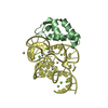
| |||||||||||||||||||||
| Unit cell |
| |||||||||||||||||||||
| Noncrystallographic symmetry (NCS) | NCS domain:
NCS oper:
|
-Components
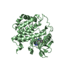
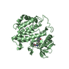
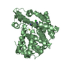
-RNA chain / Protein , 2 types, 4 molecules CDAB
| #1: RNA chain | Mass: 18693.145 Da / Num. of mol.: 2 / Fragment: RESIDUES 1051-1108 / Mutation: U1108C / Source method: obtained synthetically / Details: IN VITRO TRANSCRIBED RRNA FROM THERMOTOGA MARITIMA #2: Protein | Mass: 14980.726 Da / Num. of mol.: 2 Source method: isolated from a genetically manipulated source Details: COVALENT MERCURY LIGAND AT CYS39 / Source: (gene. exp.) Thermotoga maritima (bacteria) / Description: RECOMBINANT PROTEIN / Plasmid: PET13A / Species (production host): Escherichia coli / Production host: Escherichia coli BL21(DE3) (bacteria) / Strain (production host): BL21(DE3) / References: UniProt: P29395 |
|---|
-Non-polymers , 4 types, 177 molecules 


| #3: Chemical | ChemComp-CD /  Mass: 112.411 Da / Num. of mol.: 8 / Source method: obtained synthetically / Formula: Cd Mass: 112.411 Da / Num. of mol.: 8 / Source method: obtained synthetically / Formula: Cd#4: Chemical | ChemComp-MG /  Mass: 24.305 Da / Num. of mol.: 19 / Source method: obtained synthetically / Formula: Mg Mass: 24.305 Da / Num. of mol.: 19 / Source method: obtained synthetically / Formula: Mg#5: Chemical | ChemComp-MMC / Methylmercury Mass: 215.625 Da / Num. of mol.: 8 / Source method: obtained synthetically / Formula: CH3Hg Mass: 215.625 Da / Num. of mol.: 8 / Source method: obtained synthetically / Formula: CH3Hg#6: Water | ChemComp-HOH / | WaterMass: 18.015 Da / Num. of mol.: 142 / Source method: isolated from a natural source / Formula: H2O |
|---|
-Details
| Nonpolymer details | THERE ARE A TOTAL OF 8 METHYLMERCURY IONS IN THE STRUCTURE. THE METHYL GROUP HAS NOT BEEN MODELLED ...THERE ARE A TOTAL OF 8 METHYLMERC |
|---|
-Experimental details
-Experiment
| Experiment | Method: X-RAY DIFFRACTION / Number of used crystals: 1 |
|---|
- Sample preparation
| Crystal | Density Matthews: 3.1 Å3/Da / Density % sol: 55 % / Description: TWO WAVELENGTH HG MAD | ||||||||||||||||||||||||||||||||||||||||||||||||||||||
|---|---|---|---|---|---|---|---|---|---|---|---|---|---|---|---|---|---|---|---|---|---|---|---|---|---|---|---|---|---|---|---|---|---|---|---|---|---|---|---|---|---|---|---|---|---|---|---|---|---|---|---|---|---|---|---|
| Crystal grow | Temperature: 277 K / Method: vapor diffusion, hanging drop / pH: 8.3 Details: 25% GLYCEROL, 15% PEG 4000, 50 MM TRIS PH 7.5, 50 MM MGCL2, 20 MM CDCL2, 0.2 M KCL, 1 MM DITHIOTHREITOL, 4 DEGREES C, pH 8.3, VAPOR DIFFUSION, HANGING DROP, temperature 277K | ||||||||||||||||||||||||||||||||||||||||||||||||||||||
| Components of the solutions |
| ||||||||||||||||||||||||||||||||||||||||||||||||||||||
| Crystal | *PLUS | ||||||||||||||||||||||||||||||||||||||||||||||||||||||
| Crystal grow | *PLUS Temperature: 4 ℃ / pH: 7.5 | ||||||||||||||||||||||||||||||||||||||||||||||||||||||
| Components of the solutions | *PLUS
|
-Data collection
| Diffraction | Mean temperature: 100 K | |||||||||
|---|---|---|---|---|---|---|---|---|---|---|
| Diffraction source | Source: SYNCHROTRON / Site: NSLS / Beamline: X12C / Wavelength: 0.980,1.010 | |||||||||
| Detector | Type: BRANDEIS / Detector: CCD / Details: MIRRORS | |||||||||
| Radiation | Monochromator: SI CRYSTAL / Protocol: MAD / Monochromatic (M) / Laue (L): M / Scattering type: x-ray | |||||||||
| Radiation wavelength |
| |||||||||
| Reflection | Resolution: 2.57→20 Å / Num. all: 49313 / Num. obs: 49313 / % possible obs: 95.5 % / Observed criterion σ(I): 0 / Redundancy: 4 % / Biso Wilson estimate: 58.8 Å2 / Rsym value: 0.041 / Net I/σ(I): 20 | |||||||||
| Reflection shell | Resolution: 2.57→2.73 Å / Redundancy: 3.4 % / Mean I/σ(I) obs: 4.3 / Rsym value: 0.17 / % possible all: 86.2 | |||||||||
| Reflection | *PLUS |
- Processing
| Software |
| ||||||||||||||||||||||||||||||||||||||||||||||||||||||||||||||||||||||||||||||||
|---|---|---|---|---|---|---|---|---|---|---|---|---|---|---|---|---|---|---|---|---|---|---|---|---|---|---|---|---|---|---|---|---|---|---|---|---|---|---|---|---|---|---|---|---|---|---|---|---|---|---|---|---|---|---|---|---|---|---|---|---|---|---|---|---|---|---|---|---|---|---|---|---|---|---|---|---|---|---|---|---|---|
| Refinement | Method to determine structure: MAD / Resolution: 2.57→20 Å / Rfactor Rfree error: 0.005 / Data cutoff high absF: 1000000 / Data cutoff low absF: 0.001 / Isotropic thermal model: RESTRAINED / Cross valid method: THROUGHOUT / σ(F): 0 Details: NCS RESTRAINTS APPLIED TO RNA THROUGHOUT, NOT TO PROTEIN THE ASYMMETRIC UNIT CONTAINS TWO L11-RNA COMPLEXES. COMPLEX 1 CONSISTS OF CHAINS A AND C, AND COMPLEX 2 CONSISTS OF CHAINS B AND D. ...Details: NCS RESTRAINTS APPLIED TO RNA THROUGHOUT, NOT TO PROTEIN THE ASYMMETRIC UNIT CONTAINS TWO L11-RNA COMPLEXES. COMPLEX 1 CONSISTS OF CHAINS A AND C, AND COMPLEX 2 CONSISTS OF CHAINS B AND D. RESIDUES 1-7 AND 141 OF CHAIN A ARE DISORDERED. THE DENSITY FOR RESIDUES 8-70 OF CHAIN A WAS OF SIGNIFICANTLY LOWER QUALITY THAN THE DENSITY FOR THE REMAINDER OF THE ASYMMETRIC UNIT, AND THE QUALITY OF THE MODEL FOR THIS N-TERMINAL DOMAIN IS LOWER THAN THAT OF THE C-TERMINAL DOMAIN (RESIDUES 71-140). RESIDUES 1-70 AND 141 OF CHAIN B ARE DISORDERED. THE RNA IS NUMBERED WITH THE E. COLI NUMBERING TO FACILITATE COMPARISON WITH THE EXTENSIVE BIOCHEMICAL DATA ON THE E. COLI RNA-L11 SYSTEM. THE E. COLI RNA NUMBERING IS ALSO USED IN THE PRIMARY REFERENCE DESCRIBING THIS STRUCTURE.
| ||||||||||||||||||||||||||||||||||||||||||||||||||||||||||||||||||||||||||||||||
| Displacement parameters | Biso mean: 42.8 Å2 | ||||||||||||||||||||||||||||||||||||||||||||||||||||||||||||||||||||||||||||||||
| Refine analyze |
| ||||||||||||||||||||||||||||||||||||||||||||||||||||||||||||||||||||||||||||||||
| Refinement step | Cycle: LAST / Resolution: 2.57→20 Å
| ||||||||||||||||||||||||||||||||||||||||||||||||||||||||||||||||||||||||||||||||
| Refine LS restraints |
| ||||||||||||||||||||||||||||||||||||||||||||||||||||||||||||||||||||||||||||||||
| Refine LS restraints NCS | NCS model details: RESTRAINTS / Refine-ID: X-RAY DIFFRACTION / Rms dev Biso : 5.51 Å2 / Rms dev position: 0.07 Å / Weight Biso : 2 / Weight position: 50
| ||||||||||||||||||||||||||||||||||||||||||||||||||||||||||||||||||||||||||||||||
| LS refinement shell | Resolution: 2.57→2.73 Å / Rfactor Rfree error: 0.024 / Total num. of bins used: 6
| ||||||||||||||||||||||||||||||||||||||||||||||||||||||||||||||||||||||||||||||||
| Xplor file |
| ||||||||||||||||||||||||||||||||||||||||||||||||||||||||||||||||||||||||||||||||
| Software | *PLUS Name: X-PLOR / Version: 3.851 / Classification: refinement | ||||||||||||||||||||||||||||||||||||||||||||||||||||||||||||||||||||||||||||||||
| Refinement | *PLUS Lowest resolution: 20 Å / σ(F): 0 / % reflection Rfree: 4.9 % / Rfactor obs: 0.228 | ||||||||||||||||||||||||||||||||||||||||||||||||||||||||||||||||||||||||||||||||
| Solvent computation | *PLUS | ||||||||||||||||||||||||||||||||||||||||||||||||||||||||||||||||||||||||||||||||
| Displacement parameters | *PLUS Biso mean: 42.8 Å2 | ||||||||||||||||||||||||||||||||||||||||||||||||||||||||||||||||||||||||||||||||
| Refine LS restraints | *PLUS
| ||||||||||||||||||||||||||||||||||||||||||||||||||||||||||||||||||||||||||||||||
| LS refinement shell | *PLUS Rfactor Rfree: 0.432 / % reflection Rfree: 4.5 % / Rfactor Rwork: 0.386 |
