 Movie
Movie Controller


+ Open data

- Basic information
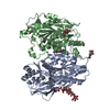
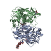
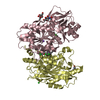
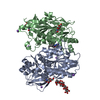
| Entry | Database: PDB / ID: 1an7 | ||||||
|---|---|---|---|---|---|---|---|
| Title | RIBOSOMAL PROTEIN S8 FROM THERMUS THERMOPHILUS | ||||||
 Components Components | RIBOSOMAL PROTEIN S8 | ||||||
 Keywords Keywords | RIBOSOMAL PROTEIN / RRNA-PROTEIN BINDING / PROTEIN-PROTEIN BINDING / THERMUS THERMOPHILUS | ||||||
| Function / homology |  Function and homology informationrRNA binding / ribosome / structural constituent of ribosome / translation / ribonucleoprotein complex / cytoplasm Function and homology informationrRNA binding / ribosome / structural constituent of ribosome / translation / ribonucleoprotein complex / cytoplasmSimilarity search - Function | ||||||
| Biological species |   Thermus thermophilus (bacteria) Thermus thermophilus (bacteria) | ||||||
| Method | X-RAY DIFFRACTION / MIR / Resolution: 2.9 Å | ||||||
 Authors Authors | Nevskaya, N. / Nikonov, S. / Al-Karadaghi, S. | ||||||
 Citation Citation | Journal: J.Mol.Biol. / Year: 1998 Title: Crystal structure of ribosomal protein S8 from Thermus thermophilus reveals a high degree of structural conservation of a specific RNA binding site. Authors: Nevskaya, N. / Tishchenko, S. / Nikulin, A. / al-Karadaghi, S. / Liljas, A. / Ehresmann, B. / Ehresmann, C. / Garber, M. / Nikonov, S. #1: Journal: Proteins / Year: 1997Title: Crystallization and Preliminary Crystallographic Analysis of Ribosomal Protein S8 from Thermus Thermophilus Authors: Tishchenko, S.V. / Vysotskaya, V.S. / Fomenkova, N.P. / Nikonov, S.V. / Ehresmann, B. / Garber, M.B. | ||||||
| History |
|
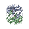
- Structure visualization
| Structure viewer | Molecule: MolmilJmol/JSmol |
|---|
- Downloads & links
-Download
| PDBx/mmCIF format | 1an7.cif.gz | 64.3 KB | Display | PDBx/mmCIF format |
|---|---|---|---|---|
| PDB format | pdb1an7.ent.gz | 49.5 KB | Display | PDB format |
| PDBx/mmJSON format | 1an7.json.gz | Tree view | PDBx/mmJSON format | |
| Others |  Other downloads Other downloads |
-Validation report
| Arichive directory | https://data.pdbj.org/pub/pdb/validation_reports/an/1an7ftp://data.pdbj.org/pub/pdb/validation_reports/an/1an7 | HTTPS FTP |
|---|
-Related structure data
| Similar structure data |
|---|


-Links
 PDBj
PDBj


- Assembly
| Deposited unit | 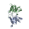
| ||||||||
|---|---|---|---|---|---|---|---|---|---|
| 1 | 
| ||||||||
| Unit cell |
| ||||||||
| Noncrystallographic symmetry (NCS) | NCS oper: (Code: given Matrix: (-0.8928, 0.0842, -0.4425), Vector : |
-Components
| #1: Protein | Mass: 15624.214 Da / Num. of mol.: 2 Source method: isolated from a genetically manipulated source Source: (gene. exp.) Thermus thermophilus (bacteria) / Strain: VK1 / Cell line: BL21 / Gene: RPS8 / Plasmid: PET-TTHS8 / Species (production host): Escherichia coli / Production host: Escherichia coli BL21(DE3) (bacteria) / Strain (production host): BL21 (DE3) / References: UniProt: P24319 |
|---|
-Experimental details
-Experiment
| Experiment | Method: X-RAY DIFFRACTION |
|---|
- Sample preparation
| Crystal | Density Matthews: 3.21 Å3/Da / Density % sol: 60 % | ||||||||||||||||||||||||||||||
|---|---|---|---|---|---|---|---|---|---|---|---|---|---|---|---|---|---|---|---|---|---|---|---|---|---|---|---|---|---|---|---|
| Crystal grow | pH: 6 / Details: pH 6.0 | ||||||||||||||||||||||||||||||
| Crystal | *PLUS | ||||||||||||||||||||||||||||||
| Crystal grow | *PLUS Temperature: 24 ℃ / Method: vapor diffusion, hanging dropDetails: Tishchenko, S.V., (1997) Proteins: Struct.,Funct., Genet., 27, 309. | ||||||||||||||||||||||||||||||
| Components of the solutions | *PLUS
|
-Data collection
| Diffraction | Mean temperature: 293 K |
|---|---|
| Diffraction source | Source: ROTATING ANODE / Type: RIGAKU RUH2R / Wavelength: 1.5418 |
| Detector | Type: MAR scanner 300 mm plate / Detector: IMAGE PLATE / Date: Feb 10, 1996 |
| Radiation | Monochromator: GRAPHITE(002) / Monochromatic (M) / Laue (L): M / Scattering type: x-ray |
| Radiation wavelength | Wavelength: 1.5418 Å / Relative weight: 1 |
| Reflection | Resolution: 2.9→20 Å / Num. obs: 8932 / % possible obs: 94.5 % / Observed criterion σ(I): 0 / Redundancy: 3.6 % / Biso Wilson estimate: 33.6 Å2 / Rmerge(I) obs: 0.051 |
| Reflection shell | Resolution: 2.9→2.95 Å / Redundancy: 3.2 % / Rmerge(I) obs: 0.019 / % possible all: 93.6 |
- Processing
| Software |
| ||||||||||||||||||||||||||||||||||||||||||||||||||||||||||||
|---|---|---|---|---|---|---|---|---|---|---|---|---|---|---|---|---|---|---|---|---|---|---|---|---|---|---|---|---|---|---|---|---|---|---|---|---|---|---|---|---|---|---|---|---|---|---|---|---|---|---|---|---|---|---|---|---|---|---|---|---|---|
| Refinement | Method to determine structure: MIR / Resolution: 2.9→8 Å
| ||||||||||||||||||||||||||||||||||||||||||||||||||||||||||||
| Displacement parameters | Biso mean: 37 Å2 | ||||||||||||||||||||||||||||||||||||||||||||||||||||||||||||
| Refinement step | Cycle: LAST / Resolution: 2.9→8 Å
| ||||||||||||||||||||||||||||||||||||||||||||||||||||||||||||
| Refine LS restraints |
| ||||||||||||||||||||||||||||||||||||||||||||||||||||||||||||
| Xplor file |
| ||||||||||||||||||||||||||||||||||||||||||||||||||||||||||||
| Software | *PLUS Name: X-PLOR / Version: 3.1 / Classification: refinement | ||||||||||||||||||||||||||||||||||||||||||||||||||||||||||||
| Refine LS restraints | *PLUS
|
