 Movie
Movie Controller

[English] 日本語
 Yorodumi
- EMDB-8315: Structure of the STRA6 receptor for retinol uptake in complex wit... -
+ Open data

- Basic information
| Entry | Database: EMDB / ID: EMD-8315 | |||||||||
|---|---|---|---|---|---|---|---|---|---|---|
| Title | Structure of the STRA6 receptor for retinol uptake in complex with calmodulin | |||||||||
 Map data Map data | Reconstruction of STRA6/CaM complex | |||||||||
 Sample Sample |
| |||||||||
 Keywords Keywords | Vitamin A /  retinol / STRA6 / membrane / MEMBRANE PROTEIN-CALCIUM BINDING PROTEIN complex retinol / STRA6 / membrane / MEMBRANE PROTEIN-CALCIUM BINDING PROTEIN complex | |||||||||
| Function / homology |  Function and homology information Function and homology informationvitamin A import into cell / The canonical retinoid cycle in rods (twilight vision) / retinol transport / retinol transmembrane transporter activity / chordate embryonic development / retinal binding / retinol binding / plasma membrane => GO:0005886 / calcium-mediated signaling / signaling receptor activity ...vitamin A import into cell / The canonical retinoid cycle in rods (twilight vision) / retinol transport / retinol transmembrane transporter activity / chordate embryonic development / retinal binding / retinol binding / plasma membrane => GO:0005886 / calcium-mediated signaling / signaling receptor activity / molecular adaptor activity / calmodulin binding / calcium ion binding / identical protein binding / plasma membraneSimilarity search - Function | |||||||||
| Biological species |  Danio rerio (zebrafish) / Danio rerio (zebrafish) /   Spodoptera frugiperda (fall armyworm) Spodoptera frugiperda (fall armyworm) | |||||||||
| Method | single particle reconstruction / cryo EM / Resolution: 3.9 Å | |||||||||
 Authors Authors | Clarke OB / Chen Y | |||||||||
 Citation Citation | Journal: Science / Year: 2016 Title: Structure of the STRA6 receptor for retinol uptake. Authors: Yunting Chen / Oliver B Clarke / Jonathan Kim / Sean Stowe / Youn-Kyung Kim / Zahra Assur / Michael Cavalier / Raquel Godoy-Ruiz / Desiree C von Alpen / Chiara Manzini / William S Blaner / ...Authors: Yunting Chen / Oliver B Clarke / Jonathan Kim / Sean Stowe / Youn-Kyung Kim / Zahra Assur / Michael Cavalier / Raquel Godoy-Ruiz / Desiree C von Alpen / Chiara Manzini / William S Blaner / Joachim Frank / Loredana Quadro / David J Weber / Lawrence Shapiro / Wayne A Hendrickson / Filippo Mancia /  Abstract: Vitamin A homeostasis is critical to normal cellular function. Retinol-binding protein (RBP) is the sole specific carrier in the bloodstream for hydrophobic retinol, the main form in which vitamin A ...Vitamin A homeostasis is critical to normal cellular function. Retinol-binding protein (RBP) is the sole specific carrier in the bloodstream for hydrophobic retinol, the main form in which vitamin A is transported. The integral membrane receptor STRA6 mediates cellular uptake of vitamin A by recognizing RBP-retinol to trigger release and internalization of retinol. We present the structure of zebrafish STRA6 determined to 3.9-angstrom resolution by single-particle cryo-electron microscopy. STRA6 has one intramembrane and nine transmembrane helices in an intricate dimeric assembly. Unexpectedly, calmodulin is bound tightly to STRA6 in a noncanonical arrangement. Residues involved with RBP binding map to an archlike structure that covers a deep lipophilic cleft. This cleft is open to the membrane, suggesting a possible mode for internalization of retinol through direct diffusion into the lipid bilayer. | |||||||||
| History |
|
- Structure visualization
| Movie |
Movie viewer |
|---|---|
| Structure viewer | EM map: SurfViewMolmilJmol/JSmol |
| Supplemental images |

- Downloads & links
-EMDB archive
| Map data | emd_8315.map.gz | 28.4 MB | EMDB map data format | |
|---|---|---|---|---|
| Header (meta data) | emd-8315-v30.xmlemd-8315.xml | 17.6 KB 17.6 KB | Display Display | EMDB header |
| FSC (resolution estimation) | emd_8315_fsc.xml | 7.1 KB | Display | FSC data file |
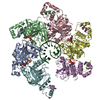
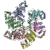
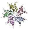
| Images | 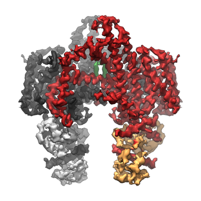 emd_8315.png emd_8315.png | 147.5 KB | ||
| Masks | emd_8315_msk_1.map | 30.5 MB | Mask map | |
| Filedesc metadata | emd-8315.cif.gz | 6.3 KB | ||
| Others | emd_8315_half_map_1.map.gzemd_8315_half_map_2.map.gz | 22.5 MB 22.5 MB | ||
| Archive directory |  http://ftp.pdbj.org/pub/emdb/structures/EMD-8315ftp://ftp.pdbj.org/pub/emdb/structures/EMD-8315 http://ftp.pdbj.org/pub/emdb/structures/EMD-8315ftp://ftp.pdbj.org/pub/emdb/structures/EMD-8315 | HTTPS FTP |
-Related structure data
| Related structure data |  5sy1MC  5k8qC M: atomic model generated by this map C: citing same article ( |
|---|---|
| Similar structure data |


-Links
| EMDB pages | EMDB (EBI/PDBe) / EMDataResource |
|---|---|
| Related items in Molecule of the Month |


-Map
| File | Download / File: emd_8315.map.gz / Format: CCP4 / Size: 30.5 MB / Type: IMAGE STORED AS FLOATING POINT NUMBER (4 BYTES) | ||||||||||||||||||||||||||||||||||||||||||||||||||||||||||||||||||||
|---|---|---|---|---|---|---|---|---|---|---|---|---|---|---|---|---|---|---|---|---|---|---|---|---|---|---|---|---|---|---|---|---|---|---|---|---|---|---|---|---|---|---|---|---|---|---|---|---|---|---|---|---|---|---|---|---|---|---|---|---|---|---|---|---|---|---|---|---|---|
| Annotation | Reconstruction of STRA6/CaM complex | ||||||||||||||||||||||||||||||||||||||||||||||||||||||||||||||||||||
| Projections & slices | Image control
Images are generated by Spider. | ||||||||||||||||||||||||||||||||||||||||||||||||||||||||||||||||||||
| Voxel size | X=Y=Z: 1.255 Å | ||||||||||||||||||||||||||||||||||||||||||||||||||||||||||||||||||||
| Density |
| ||||||||||||||||||||||||||||||||||||||||||||||||||||||||||||||||||||
| Symmetry | Space group: 1 | ||||||||||||||||||||||||||||||||||||||||||||||||||||||||||||||||||||
| Details | EMDB XML:
CCP4 map header:
| ||||||||||||||||||||||||||||||||||||||||||||||||||||||||||||||||||||
 Z (Sec.)
 Y (Row.)
 X (Col.)


-Supplemental data
-Mask #1
| File | emd_8315_msk_1.map | ||||||||||||
|---|---|---|---|---|---|---|---|---|---|---|---|---|---|
| Projections & Slices |
| ||||||||||||
| Density Histograms |


-Half map: Reconstruction of STRA6/CaM complex, half map 1
| File | emd_8315_half_map_1.map | ||||||||||||
|---|---|---|---|---|---|---|---|---|---|---|---|---|---|
| Annotation | Reconstruction of STRA6/CaM complex, half map 1 | ||||||||||||
| Projections & Slices |
| ||||||||||||
| Density Histograms |


-Half map: Reconstruction of STRA6/CaM complex, half map 2
| File | emd_8315_half_map_2.map | ||||||||||||
|---|---|---|---|---|---|---|---|---|---|---|---|---|---|
| Annotation | Reconstruction of STRA6/CaM complex, half map 2 | ||||||||||||
| Projections & Slices |
| ||||||||||||
| Density Histograms |


- Sample components
-Entire : Complex of zebrafish (D. rerio) STRA6 with copurified calmodulin ...
| Entire | Name: Complex of zebrafish (D. rerio) STRA6 with copurified calmodulin reconstituted in amphipol A8-35 |
|---|---|
| Components |
|
-Supramolecule #1: Complex of zebrafish (D. rerio) STRA6 with copurified calmodulin ...
| Supramolecule | Name: Complex of zebrafish (D. rerio) STRA6 with copurified calmodulin reconstituted in amphipol A8-35 type: complex / ID: 1 / Parent: 0 / Macromolecule list: #1-#2 |
|---|---|
| Source (natural) | Organism: Danio rerio (zebrafish) |
-Macromolecule #1: Calmodulin
| Macromolecule | Name: Calmodulin / type: protein_or_peptide / ID: 1 / Number of copies: 2 / Enantiomer: LEVO |
|---|---|
| Source (natural) | Organism: Spodoptera frugiperda (fall armyworm) |
| Molecular weight | Theoretical: 16.82552 KDa |
| Sequence | String: MADQLTEEQI AEFKEAFSLF DKDGDGTITT KELGTVMRSL GQNPTEAELQ DMINEVDADG NGTIDFPEFL TMMARKMKDT DSEEEIREA FRVFDKDGNG FISAAELRHV MTNLGEKLTD EEVDEMIREA DIDGDGQVNY EEFVTMMTSK |
-Macromolecule #2: STRA6
| Macromolecule | Name: STRA6 / type: protein_or_peptide / ID: 2 / Number of copies: 2 / Enantiomer: LEVO |
|---|---|
| Source (natural) | Organism: Danio rerio (zebrafish) |
| Molecular weight | Theoretical: 75.551523 KDa |
| Recombinant expression | Organism: Spodoptera frugiperda (fall armyworm) |
| Sequence | String: MSAETVNNYD YSDWYENAAP TKAPVEVIPP CDPTADEGLF HICIAAISLV VMLVLAILAR RQKLSDNQRG LTGLLSPVNF LDHTQHKGL AVAVYGVLFC KLVGMVLSHH PLPFTKEVAN KEFWMILALL YYPTLYYPLL ACGTLHNKVG YVLGSLLSWT H FGILVWQK ...String: MSAETVNNYD YSDWYENAAP TKAPVEVIPP CDPTADEGLF HICIAAISLV VMLVLAILAR RQKLSDNQRG LTGLLSPVNF LDHTQHKGL AVAVYGVLFC KLVGMVLSHH PLPFTKEVAN KEFWMILALL YYPTLYYPLL ACGTLHNKVG YVLGSLLSWT H FGILVWQK VDCPKTPQIY KYYALFGSLP QIACLAFLSF QYPLLLFKGL QNTETANASE DLSSSYYRDY VKKILKKKKP TK ISSSTSK PKLFDRLRDA VKSYIYTPED VFRFPLKLAI SVVVAFIALY QMALLLISGV LPTLHIVRRG VDENIAFLLA GFN IILSND RQEVVRIVVY YLWCVEICYV SAVTLSCLVN LLMLMRSMVL HRSNLKGLYR GDSLNVFNCH RSIRPSRPAL VCWM GFTSY QAAFLCLGMA IQTLVFFICI LFAVFLIIIP ILWGTNLMLF HIIGNLWPFW LTLVLAALIQ HVASRFLFIR KDGGT RDLN NRGSLFLLSY ILFLVNVMIG VVLGIWRVVI TALFNIVHLG RLDISLLNRN VEAFDPGYRC YSHYLKIEVS QSHPVM KAF CGLLLQSSGQ DGLSAQRIRD AEEGIQLVQQ EKKQNKVSNA KRARAHWQLL YTLVNNPSLV GSRKHFQCQS SESFING AL SRTSKEGSKK DGSVKEPNKE AESAAASN UniProtKB: Receptor for retinol uptake stra6 |
-Macromolecule #3: CALCIUM ION
| Macromolecule | Name: CALCIUM ION / type: ligand / ID: 3 / Number of copies: 8 / Formula: CA |
|---|---|
| Molecular weight | Theoretical: 40.078 Da |
-Macromolecule #4: CHOLESTEROL
| Macromolecule | Name: CHOLESTEROL / type: ligand / ID: 4 / Number of copies: 2 / Formula: CLR |
|---|---|
| Molecular weight | Theoretical: 386.654 Da |
| Chemical component information |  ChemComp-CLR: |
-Experimental details
-Structure determination
| Method | cryo EM |
|---|---|
 Processing Processing | single particle reconstruction |
| Aggregation state | particle |
-Sample preparation
| Concentration | 0.6 mg/mL |
|---|---|
| Buffer | pH: 7 |
| Grid | Model: Quantifoil, UltrAuFoil, R1.2/1.3 / Material: GOLD / Mesh: 400 / Pretreatment - Type: PLASMA CLEANING |
| Vitrification | Cryogen name: ETHANE / Chamber humidity: 95 % / Chamber temperature: 277 K / Instrument: FEI VITROBOT MARK IV Details: 3 uL sample applied, 3-4 second blot time, 30 second wait time, blot force 3, grid blotted from both sides, plunged into liquid ethane (FEI VITROBOT MARK IV).. |
- Electron microscopy
| Microscope | FEI TECNAI F30 |
|---|---|
| Electron beam | Acceleration voltage: 300 kV / Electron source: FIELD EMISSION GUN |
| Electron optics | Illumination mode: FLOOD BEAM / Imaging mode: BRIGHT FIELDBright-field microscopy |
| Image recording | Film or detector model: GATAN K2 SUMMIT (4k x 4k) / Detector mode: COUNTING / Average electron dose: 100.0 e/Å2 |
| Experimental equipment |  Model: Tecnai F30 / Image courtesy: FEI Company |
-Image processing
| Startup model | Type of model: OTHER Details: An initial model was generated using e2initialmodel.py starting from 2D class averages created in Relion 1.3. |
|---|---|
| Initial angle assignment | Type: PROJECTION MATCHING |
| Final angle assignment | Type: PROJECTION MATCHING / Software - Name: RELION (ver. 1.3) |
| Final reconstruction | Applied symmetry - Point group: C2 (2 fold cyclic) / Resolution.type: BY AUTHOR / Resolution: 3.9 Å / Resolution method: FSC 0.143 CUT-OFF / Software - Name: RELION (ver. 1.3) / Number images used: 56615 |
| FSC plot (resolution estimation) |  |
-Atomic model buiding 1
| Refinement | Protocol: AB INITIO MODEL |
|---|---|
| Output model | PDB-5sy1: |
