 Movie
Movie Controller


+ Open data

- Basic information
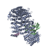
| Entry | Database: EMDB / ID: EMD-8295 | |||||||||
|---|---|---|---|---|---|---|---|---|---|---|
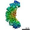
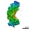
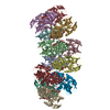
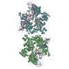
| Title | Structure of 2x duplex DNA target-bound Cascade/I-C | |||||||||
 Map data Map data | Structure of 2x duplex DNA target-bound Cascade/I-C | |||||||||
 Sample Sample |
| |||||||||
| Biological species |   Desulfovibrio vulgaris (bacteria) Desulfovibrio vulgaris (bacteria) | |||||||||
| Method | single particle reconstruction / cryo EM / Resolution: 8.8 Å | |||||||||
 Authors Authors | Hochstrasser JL / Taylor DW / Kornfeld JK / Nogales E / Doudna JA | |||||||||
| Funding support |  United States, 1 items United States, 1 items
| |||||||||
 Citation Citation | Journal: Mol Cell / Year: 2016 Title: DNA Targeting by a Minimal CRISPR RNA-Guided Cascade. Authors: Megan L Hochstrasser / David W Taylor / Jack E Kornfeld / Eva Nogales / Jennifer A Doudna / Abstract: Bacteria employ surveillance complexes guided by CRISPR (clustered, regularly interspaced, short palindromic repeats) RNAs (crRNAs) to target foreign nucleic acids for destruction. Although most ...Bacteria employ surveillance complexes guided by CRISPR (clustered, regularly interspaced, short palindromic repeats) RNAs (crRNAs) to target foreign nucleic acids for destruction. Although most type I and type III CRISPR systems require four or more distinct proteins to form multi-subunit surveillance complexes, the type I-C systems use just three proteins to achieve crRNA maturation and double-stranded DNA target recognition. We show that each protein plays multiple functional and structural roles: Cas5c cleaves pre-crRNAs and recruits Cas7 to position the RNA guide for DNA binding and unwinding by Cas8c. Cryoelectron microscopy reconstructions of free and DNA-bound forms of the Cascade/I-C surveillance complex reveal conformational changes that enable R-loop formation with distinct positioning of each DNA strand. This streamlined type I-C system explains how CRISPR pathways can evolve compact structures that retain full functionality as RNA-guided DNA capture platforms. | |||||||||
| History |
|
- Structure visualization
| Movie |
 Movie viewer Movie viewer |
|---|---|
| Structure viewer | EM map: SurfViewMolmilJmol/JSmol |
| Supplemental images |

- Downloads & links
-EMDB archive
| Map data | emd_8295.map.gz | 14.6 MB | EMDB map data format | |
|---|---|---|---|---|
| Header (meta data) | emd-8295-v30.xmlemd-8295.xml | 12 KB 12 KB | Display Display | EMDB header |
| Images |  emd_8295.png emd_8295.png | 94.1 KB | ||
| Archive directory |  http://ftp.pdbj.org/pub/emdb/structures/EMD-8295ftp://ftp.pdbj.org/pub/emdb/structures/EMD-8295 http://ftp.pdbj.org/pub/emdb/structures/EMD-8295ftp://ftp.pdbj.org/pub/emdb/structures/EMD-8295 | HTTPS FTP |
-Related structure data


-Links
| EMDB pages | EMDB (EBI/PDBe) / EMDataResource |
|---|
-Map
| File | Download / File: emd_8295.map.gz / Format: CCP4 / Size: 15.6 MB / Type: IMAGE STORED AS FLOATING POINT NUMBER (4 BYTES) | ||||||||||||||||||||||||||||||||||||||||||||||||||||||||||||||||||||
|---|---|---|---|---|---|---|---|---|---|---|---|---|---|---|---|---|---|---|---|---|---|---|---|---|---|---|---|---|---|---|---|---|---|---|---|---|---|---|---|---|---|---|---|---|---|---|---|---|---|---|---|---|---|---|---|---|---|---|---|---|---|---|---|---|---|---|---|---|---|
| Annotation | Structure of 2x duplex DNA target-bound Cascade/I-C | ||||||||||||||||||||||||||||||||||||||||||||||||||||||||||||||||||||
| Voxel size | X=Y=Z: 2.04 Å | ||||||||||||||||||||||||||||||||||||||||||||||||||||||||||||||||||||
| Density |
| ||||||||||||||||||||||||||||||||||||||||||||||||||||||||||||||||||||
| Symmetry | Space group: 1 | ||||||||||||||||||||||||||||||||||||||||||||||||||||||||||||||||||||
| Details | EMDB XML:
CCP4 map header:
| ||||||||||||||||||||||||||||||||||||||||||||||||||||||||||||||||||||
-Supplemental data
- Sample components
-Entire : 2x duplex DNA target-bound Cascade
| Entire | Name: 2x duplex DNA target-bound Cascade |
|---|---|
| Components |
|
-Supramolecule #1: 2x duplex DNA target-bound Cascade
| Supramolecule | Name: 2x duplex DNA target-bound Cascade / type: complex / ID: 1 / Parent: 0 / Details: apo-Cascade/I-C bound to 75 bp duplex DNA |
|---|---|
| Source (natural) | Organism: Desulfovibrio vulgaris (bacteria) |
| Recombinant expression | Organism: Escherichia coli (E. coli) |
-Experimental details
-Structure determination
| Method | cryo EM |
|---|---|
 Processing Processing | single particle reconstruction |
| Aggregation state | particle |
-Sample preparation
| Concentration | 0.25 mg/mL | ||||||||||||
|---|---|---|---|---|---|---|---|---|---|---|---|---|---|
| Buffer | pH: 7.5 Component:
| ||||||||||||
| Grid | Model: Protochips C-flat 4/2 / Material: COPPER / Support film - Material: CARBON / Support film - topology: CONTINUOUS / Pretreatment - Type: PLASMA CLEANING | ||||||||||||
| Vitrification | Cryogen name: ETHANE / Chamber humidity: 100 % / Chamber temperature: 277 K / Instrument: FEI VITROBOT MARK IV | ||||||||||||
| Details | The sample was monodisperse. |
- Electron microscopy
| Microscope | FEI TITAN KRIOS |
|---|---|
| Electron beam | Acceleration voltage: 300 kV / Electron source: FIELD EMISSION GUN |
| Electron optics | Illumination mode: FLOOD BEAM / Imaging mode: BRIGHT FIELDBright-field microscopy / Nominal defocus max: 4.5 µm / Nominal defocus min: 2.0 µm / Nominal magnification: 27000 |
| Sample stage | Specimen holder model: FEI TITAN KRIOS AUTOGRID HOLDER |
| Image recording | Film or detector model: GATAN K2 SUMMIT (4k x 4k) / Detector mode: COUNTING / Number grids imaged: 1 / Number real images: 4200 / Average exposure time: 6.0 sec. / Average electron dose: 48.0 e/Å2 |
| Experimental equipment |  Model: Titan Krios / Image courtesy: FEI Company |
-Image processing
| CTF correction | Software - Name: CTFFIND (ver. 3) |
|---|---|
| Startup model | Type of model: OTHER / Details: Negative stain model of complex |
| Initial angle assignment | Type: PROJECTION MATCHING / Software - Name: EMAN (ver. 2) |
| Final 3D classification | Number classes: 3 / Software - Name: RELION (ver. 1.4) |
| Final angle assignment | Type: PROJECTION MATCHING / Software - Name: EMAN (ver. 2) |
| Final reconstruction | Resolution.type: BY AUTHOR / Resolution: 8.8 Å / Resolution method: FSC 0.143 CUT-OFF / Software - Name: RELION (ver. 1.4) / Number images used: 17000 |
