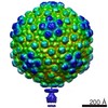
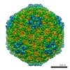
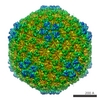
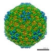
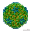
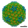
Journal: mBio / Year: 2016 Title: Localization of the Houdinisome (Ejection Proteins) inside the Bacteriophage P22 Virion by Bubblegram Imaging. Authors: Weimin Wu / Justin C Leavitt / Naiqian Cheng / Eddie B Gilcrease / Tina Motwani / Carolyn M Teschke / Sherwood R Casjens / Alasdair C Steven / Abstract: The P22 capsid is a T=7 icosahedrally symmetric protein shell with a portal protein dodecamer at one 5-fold vertex. Extending outwards from that vertex is a short tail, and putatively extending ...The P22 capsid is a T=7 icosahedrally symmetric protein shell with a portal protein dodecamer at one 5-fold vertex. Extending outwards from that vertex is a short tail, and putatively extending inwards is a 15-nm-long α-helical barrel formed by the C-terminal domains of portal protein subunits. In addition to the densely packed genome, the capsid contains three "ejection proteins" (E-proteins [gp7, gp16, and gp20]) destined to exit from the tightly sealed capsid during the process of DNA delivery into target cells. We estimated their copy numbers by quantitative SDS-PAGE as approximately 12 molecules per virion of gp16 and gp7 and 30 copies of gp20. To localize them, we used bubblegram imaging, an adaptation of cryo-electron microscopy in which gaseous bubbles induced in proteins by prolonged irradiation are used to map the proteins' locations. We applied this technique to wild-type P22, a triple mutant lacking all three E-proteins, and three mutants each lacking one E-protein. We conclude that all three E-proteins are loosely clustered around the portal axis, in the region displaced radially inwards from the portal crown. The bubblegram data imply that approximately half of the α-helical barrel seen in the portal crystal structure is disordered in the mature virion, and parts of the disordered region present binding sites for E-proteins. Thus positioned, the E-proteins are strategically placed to pass down the shortened barrel and through the portal ring and the tail, as they exit from the capsid during an infection. IMPORTANCE: While it has long been appreciated that capsids serve as delivery vehicles for viral genomes, there is now growing awareness that viruses also deliver proteins into their host cells. P22 ...IMPORTANCE: While it has long been appreciated that capsids serve as delivery vehicles for viral genomes, there is now growing awareness that viruses also deliver proteins into their host cells. P22 has three such proteins (ejection proteins [E-proteins]), whose initial locations in the virion have remained unknown despite their copious amounts (total of 2.5 MDa). This study succeeded in localizing them by the novel technique of bubblegram imaging. The P22 E-proteins are seen to be distributed around the orifice of the portal barrel. Interestingly, this barrel, 15 nm long in a crystal structure, is only about half as long in situ: the remaining, disordered, portion appears to present binding sites for E-proteins. These observations document a spectacular example of a regulatory order-disorder transition in a supramolecular system and demonstrate the potential of bubblegram imaging to map the components of other viruses as well as cellular complexes.
History
Deposition
Jun 24, 2016
-
Header (metadata) release
Jul 27, 2016
-
Map release
Jul 27, 2016
-
Update
Nov 1, 2023
-
Current status
Nov 1, 2023
Processing site: RCSB / Status: Released
-
Structure visualization
Movie
Surface view with section colored by density value
In the structure databanks used in Yorodumi, some data are registered as the other names, "COVID-19 virus" and "2019-nCoV". Here are the details of the virus and the list of structure data.
Jan 31, 2019. EMDB accession codes are about to change! (news from PDBe EMDB page)
EMDB accession codes are about to change! (news from PDBe EMDB page)
The allocation of 4 digits for EMDB accession codes will soon come to an end. Whilst these codes will remain in use, new EMDB accession codes will include an additional digit and will expand incrementally as the available range of codes is exhausted. The current 4-digit format prefixed with “EMD-” (i.e. EMD-XXXX) will advance to a 5-digit format (i.e. EMD-XXXXX), and so on. It is currently estimated that the 4-digit codes will be depleted around Spring 2019, at which point the 5-digit format will come into force.
The EM Navigator/Yorodumi systems omit the EMD- prefix.
Related info.:Q: What is EMD? / ID/Accession-code notation in Yorodumi/EM Navigator
Yorodumi is a browser for structure data from EMDB, PDB, SASBDB, etc.
This page is also the successor to EM Navigator detail page, and also detail information page/front-end page for Omokage search.
The word "yorodu" (or yorozu) is an old Japanese word meaning "ten thousand". "mi" (miru) is to see.
Related info.:EMDB / PDB / SASBDB / Comparison of 3 databanks / Yorodumi Search / Aug 31, 2016. New EM Navigator & Yorodumi / Yorodumi Papers / Jmol/JSmol / Function and homology information / Changes in new EM Navigator and Yorodumi
 Movie
Movie Controller


 Open data
 Basic information
 Map data
 Sample
 Keywords
 DNA packaging /
 Authors
 Citation
 Structure visualization
 Movie viewer
 Downloads & links
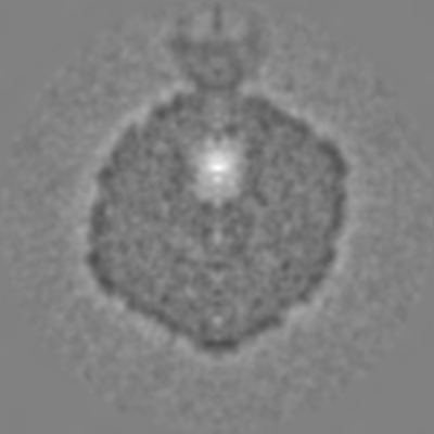 emd_8261.png
 http://ftp.pdbj.org/pub/emdb/structures/EMD-8261

 Sample components
 Processing
 Electron microscopy