 Movie
Movie Controller

[English] 日本語
 Yorodumi
- EMDB-8243: Near-atomic resolution cryo-EM reconstruction of HPV16 complexed ... -
+ Open data

- Basic information
| Entry | Database: EMDB / ID: EMD-8243 | |||||||||
|---|---|---|---|---|---|---|---|---|---|---|
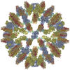
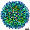
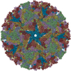
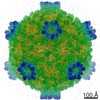
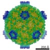
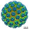
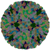
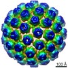
| Title | Near-atomic resolution cryo-EM reconstruction of HPV16 complexed with Fab V5 | |||||||||
 Map data Map data | HPV16 complexed with Fab V5 | |||||||||
 Sample Sample | HPV16 complexed with Fab V5 != Human papillomavirus type 16 HPV16 complexed with Fab V5
| |||||||||
| Function / homology |  Function and homology information Function and homology informationT=7 icosahedral viral capsid / endocytosis involved in viral entry into host cell / host cell nucleus / structural molecule activity / virion attachment to host cell Similarity search - Function | |||||||||
| Biological species |   Human papillomavirus type 16 Human papillomavirus type 16 | |||||||||
| Method | single particle reconstruction / cryo EM / Resolution: 4.7 Å | |||||||||
 Authors Authors | Guan J / Bywaters SM / Brendle SA / Ashley RE / Makhov AM / Conway JF / Christensen ND / Hafenstein S | |||||||||
 Citation Citation | Journal: Viruses / Year: 2017 Title: High-Resolution Structure Analysis of Antibody V5 and U4 Conformational Epitopes on Human Papillomavirus 16. Authors: Jian Guan / Stephanie M Bywaters / Sarah A Brendle / Robert E Ashley / Alexander M Makhov / James F Conway / Neil D Christensen / Susan Hafenstein /  Abstract: Cancers attributable to human papillomavirus (HPV) place a huge burden on the health of both men and women. The current commercial vaccines are genotype specific and provide little therapeutic ...Cancers attributable to human papillomavirus (HPV) place a huge burden on the health of both men and women. The current commercial vaccines are genotype specific and provide little therapeutic benefit to patients with existing HPV infections. Identifying the conformational epitopes on the virus capsid supports the development of improved recombinant vaccines to maximize long-term protection against multiple types of HPV. Fragments of antibody (Fab) digested from the neutralizing monoclonal antibodies H16.V5 (V5) and H16.U4 (U4) were bound to HPV16 capsids and the structures of the two virus-Fab complexes were solved to near atomic resolution using cryo-electron microscopy. The structures reveal virus conformational changes, the Fab-binding mode to the capsid, the residues comprising the epitope and indicate a potential interaction of U4 with the minor structural protein, L2. Competition enzyme-linked immunosorbent assay (ELISA) showed V5 outcompetes U4 when added sequentially, demonstrating a steric interference even though the footprints do not overlap. Combined with our previously reported immunological and structural results, we propose that the virus may initiate host entry through an interaction between the icosahedral five-fold vertex of the capsid and receptors on the host cell. The highly detailed epitopes identified for the two antibodies provide a framework for continuing biochemical, genetic and biophysical studies. | |||||||||
| History |
|
- Structure visualization
| Movie |
Movie viewer |
|---|---|
| Structure viewer | EM map: SurfViewMolmilJmol/JSmol |
| Supplemental images |

- Downloads & links
-EMDB archive
| Map data | emd_8243.map.gz | 503.9 MB | EMDB map data format | |
|---|---|---|---|---|
| Header (meta data) | emd-8243-v30.xmlemd-8243.xml | 8.5 KB 8.5 KB | Display Display | EMDB header |
| Images | 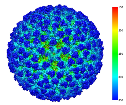 emd_8243.png emd_8243.png | 321.3 KB | ||
| Archive directory |  http://ftp.pdbj.org/pub/emdb/structures/EMD-8243ftp://ftp.pdbj.org/pub/emdb/structures/EMD-8243 http://ftp.pdbj.org/pub/emdb/structures/EMD-8243ftp://ftp.pdbj.org/pub/emdb/structures/EMD-8243 | HTTPS FTP |
-Related structure data
| Related structure data |  6bt3MC  7136C  6bspC  8245 M: atomic model generated by this map C: citing same article ( |
|---|---|
| Similar structure data |


-Links
| EMDB pages | EMDB (EBI/PDBe) / EMDataResource |
|---|---|
| Related items in Molecule of the Month |


-Map
| File | Download / File: emd_8243.map.gz / Format: CCP4 / Size: 536.4 MB / Type: IMAGE STORED AS FLOATING POINT NUMBER (4 BYTES) | ||||||||||||||||||||||||||||||||||||||||||||||||||||||||||||
|---|---|---|---|---|---|---|---|---|---|---|---|---|---|---|---|---|---|---|---|---|---|---|---|---|---|---|---|---|---|---|---|---|---|---|---|---|---|---|---|---|---|---|---|---|---|---|---|---|---|---|---|---|---|---|---|---|---|---|---|---|---|
| Annotation | HPV16 complexed with Fab V5 | ||||||||||||||||||||||||||||||||||||||||||||||||||||||||||||
| Projections & slices | Image control
Images are generated by Spider. | ||||||||||||||||||||||||||||||||||||||||||||||||||||||||||||
| Voxel size | X=Y=Z: 1.75 Å | ||||||||||||||||||||||||||||||||||||||||||||||||||||||||||||
| Density |
| ||||||||||||||||||||||||||||||||||||||||||||||||||||||||||||
| Symmetry | Space group: 1 | ||||||||||||||||||||||||||||||||||||||||||||||||||||||||||||
| Details | EMDB XML:
CCP4 map header:
| ||||||||||||||||||||||||||||||||||||||||||||||||||||||||||||
 Z (Sec.)
 Y (Row.)
 X (Col.)


-Supplemental data
- Sample components
-Entire : HPV16 complexed with Fab V5
| Entire | Name: HPV16 complexed with Fab V5 |
|---|---|
| Components |
|
-Supramolecule #1: Human papillomavirus type 16
| Supramolecule | Name: Human papillomavirus type 16 / type: virus / ID: 1 / Parent: 0 / NCBI-ID: 333760 / Sci species name: Human papillomavirus type 16 / Virus type: VIRUS-LIKE PARTICLE / Virus isolate: SEROCOMPLEX / Virus enveloped: No / Virus empty: No |
|---|---|
| Host (natural) | Organism:  Homo sapiens (human) Homo sapiens (human) |
-Experimental details
-Structure determination
| Method | cryo EM |
|---|---|
 Processing Processing | single particle reconstruction |
| Aggregation state | particle |
-Sample preparation
| Buffer | pH: 7.4 |
|---|---|
| Vitrification | Cryogen name: ETHANE |
- Electron microscopy
| Microscope | FEI POLARA 300 |
|---|---|
| Electron beam | Acceleration voltage: 300 kV / Electron source: FIELD EMISSION GUN |
| Electron optics | Illumination mode: SPOT SCAN / Imaging mode: BRIGHT FIELDBright-field microscopy |
| Image recording | Film or detector model: FEI FALCON II (4k x 4k) / Average electron dose: 7.0 e/Å2 |
| Experimental equipment |  Model: Tecnai Polara / Image courtesy: FEI Company |
-Image processing
| Startup model | Type of model: OTHER / Details: Auto3dem RMC |
|---|---|
| Initial angle assignment | Type: COMMON LINE |
| Final angle assignment | Type: COMMON LINE |
| Final reconstruction | Resolution.type: BY AUTHOR / Resolution: 4.7 Å / Resolution method: FSC 0.143 CUT-OFF / Number images used: 17612 |
