 Movie
Movie Controller

[English] 日本語
 Yorodumi
- EMDB-8150: Structure and dynamics of single-isoform recombinant neuronal hum... -
+ Open data

- Basic information
| Entry | Database: EMDB / ID: EMD-8150 | ||||||||||||
|---|---|---|---|---|---|---|---|---|---|---|---|---|---|
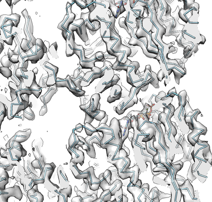
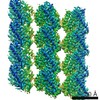
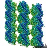
| Title | Structure and dynamics of single-isoform recombinant neuronal human tubulin | ||||||||||||
 Map data Map data | None | ||||||||||||
 Sample Sample |
| ||||||||||||
 Keywords Keywords |  microtubules / tubulin / single isoform / recombinant / dynamic instability / STRUCTURAL PROTEIN microtubules / tubulin / single isoform / recombinant / dynamic instability / STRUCTURAL PROTEIN | ||||||||||||
| Function / homology |  Function and homology informationnetrin receptor binding / Post-chaperonin tubulin folding pathway / axonemal microtubule / Carboxyterminal post-translational modifications of tubulin / dorsal root ganglion development / Microtubule-dependent trafficking of connexons from Golgi to the plasma membrane / Cilium Assembly / organelle transport along microtubule / glial cell differentiation / cytoskeleton-dependent intracellular transport ...netrin receptor binding / Post-chaperonin tubulin folding pathway / axonemal microtubule / Carboxyterminal post-translational modifications of tubulin / dorsal root ganglion development / Microtubule-dependent trafficking of connexons from Golgi to the plasma membrane / Cilium Assembly / organelle transport along microtubule / glial cell differentiation / cytoskeleton-dependent intracellular transport / forebrain morphogenesis / Intraflagellar transport / Sealing of the nuclear envelope (NE) by ESCRT-III / neuron projection arborization / Gap junction assembly / Formation of tubulin folding intermediates by CCT/TriC / cerebellar cortex morphogenesis / dentate gyrus development / COPI-independent Golgi-to-ER retrograde traffic / pyramidal neuron differentiation / Prefoldin mediated transfer of substrate to CCT/TriC / Kinesins / Assembly and cell surface presentation of NMDA receptors / centrosome cycle / COPI-dependent Golgi-to-ER retrograde traffic / motor behavior / response to L-glutamate / smoothened signaling pathway / regulation of synapse organization / intercellular bridge / locomotory exploration behavior / startle response / microtubule polymerization / Recycling pathway of L1 / RHO GTPases activate IQGAPs / Hedgehog 'off' state / response to tumor necrosis factor / microtubule-based process / COPI-mediated anterograde transport / Activation of AMPK downstream of NMDARs / response to mechanical stimulus / Mitotic Prometaphase / homeostasis of number of cells within a tissue / EML4 and NUDC in mitotic spindle formation / condensed chromosome / Loss of Nlp from mitotic centrosomes / Loss of proteins required for interphase microtubule organization from the centrosome / Recruitment of mitotic centrosome proteins and complexes / Resolution of Sister Chromatid Cohesion / Recruitment of NuMA to mitotic centrosomes / HSP90 chaperone cycle for steroid hormone receptors (SHR) in the presence of ligand / Anchoring of the basal body to the plasma membrane / cellular response to calcium ion / MHC class II antigen presentation / adult locomotory behavior / AURKA Activation by TPX2 / filopodium / cell periphery / RHO GTPases Activate Formins / Translocation of SLC2A4 (GLUT4) to the plasma membrane / axon guidance / synapse organization / peptide binding / Hydrolases; Acting on acid anhydrides; Acting on GTP to facilitate cellular and subcellular movement / intracellular protein transport / neuron migration / neuromuscular junction / visual learning / PKR-mediated signaling / structural constituent of cytoskeleton / cytoplasmic ribonucleoprotein granule / mitotic spindle / cerebral cortex development / memory / microtubule cytoskeleton organization / recycling endosome / Aggrephagy / HCMV Early Events / Separation of Sister Chromatids / The role of GTSE1 in G2/M progression after G2 checkpoint / microtubule cytoskeleton / Regulation of PLK1 Activity at G2/M Transition / mitotic cell cycle / lamellipodium / gene expression / growth cone / neuron apoptotic process / microtubule / hydrolase activity / protein heterodimerization activity / cell division / axon / GTPase activity / dendrite / neuronal cell body / protein-containing complex binding / structural molecule activity / GTP binding / extracellular exosome / identical protein binding Function and homology informationnetrin receptor binding / Post-chaperonin tubulin folding pathway / axonemal microtubule / Carboxyterminal post-translational modifications of tubulin / dorsal root ganglion development / Microtubule-dependent trafficking of connexons from Golgi to the plasma membrane / Cilium Assembly / organelle transport along microtubule / glial cell differentiation / cytoskeleton-dependent intracellular transport ...netrin receptor binding / Post-chaperonin tubulin folding pathway / axonemal microtubule / Carboxyterminal post-translational modifications of tubulin / dorsal root ganglion development / Microtubule-dependent trafficking of connexons from Golgi to the plasma membrane / Cilium Assembly / organelle transport along microtubule / glial cell differentiation / cytoskeleton-dependent intracellular transport / forebrain morphogenesis / Intraflagellar transport / Sealing of the nuclear envelope (NE) by ESCRT-III / neuron projection arborization / Gap junction assembly / Formation of tubulin folding intermediates by CCT/TriC / cerebellar cortex morphogenesis / dentate gyrus development / COPI-independent Golgi-to-ER retrograde traffic / pyramidal neuron differentiation / Prefoldin mediated transfer of substrate to CCT/TriC / Kinesins / Assembly and cell surface presentation of NMDA receptors / centrosome cycle / COPI-dependent Golgi-to-ER retrograde traffic / motor behavior / response to L-glutamate / smoothened signaling pathway / regulation of synapse organization / intercellular bridge / locomotory exploration behavior / startle response / microtubule polymerization / Recycling pathway of L1 / RHO GTPases activate IQGAPs / Hedgehog 'off' state / response to tumor necrosis factor / microtubule-based process / COPI-mediated anterograde transport / Activation of AMPK downstream of NMDARs / response to mechanical stimulus / Mitotic Prometaphase / homeostasis of number of cells within a tissue / EML4 and NUDC in mitotic spindle formation / condensed chromosome / Loss of Nlp from mitotic centrosomes / Loss of proteins required for interphase microtubule organization from the centrosome / Recruitment of mitotic centrosome proteins and complexes / Resolution of Sister Chromatid Cohesion / Recruitment of NuMA to mitotic centrosomes / HSP90 chaperone cycle for steroid hormone receptors (SHR) in the presence of ligand / Anchoring of the basal body to the plasma membrane / cellular response to calcium ion / MHC class II antigen presentation / adult locomotory behavior / AURKA Activation by TPX2 / filopodium / cell periphery / RHO GTPases Activate Formins / Translocation of SLC2A4 (GLUT4) to the plasma membrane / axon guidance / synapse organization / peptide binding / Hydrolases; Acting on acid anhydrides; Acting on GTP to facilitate cellular and subcellular movement / intracellular protein transport / neuron migration / neuromuscular junction / visual learning / PKR-mediated signaling / structural constituent of cytoskeleton / cytoplasmic ribonucleoprotein granule / mitotic spindle / cerebral cortex development / memory / microtubule cytoskeleton organization / recycling endosome / Aggrephagy / HCMV Early Events / Separation of Sister Chromatids / The role of GTSE1 in G2/M progression after G2 checkpoint / microtubule cytoskeleton / Regulation of PLK1 Activity at G2/M Transition / mitotic cell cycle / lamellipodium / gene expression / growth cone / neuron apoptotic process / microtubule / hydrolase activity / protein heterodimerization activity / cell division / axon / GTPase activity / dendrite / neuronal cell body / protein-containing complex binding / structural molecule activity / GTP binding / extracellular exosome / identical protein bindingSimilarity search - Function | ||||||||||||
| Biological species |  Homo sapiens (human) Homo sapiens (human) | ||||||||||||
| Method | helical reconstruction / cryo EM / Resolution: 4.0 Å | ||||||||||||
 Authors Authors | Vemu A / Atherton J | ||||||||||||
| Funding support |  United Kingdom, United Kingdom,  United States, 3 items United States, 3 items
| ||||||||||||
 Citation Citation | Journal: J Biol Chem / Year: 2016 Title: Structure and Dynamics of Single-isoform Recombinant Neuronal Human Tubulin. Authors: Annapurna Vemu / Joseph Atherton / Jeffrey O Spector / Agnieszka Szyk / Carolyn A Moores / Antonina Roll-Mecak / Abstract: Microtubules are polymers that cycle stochastically between polymerization and depolymerization, i.e. they exhibit "dynamic instability." This behavior is crucial for cell division, motility, and ...Microtubules are polymers that cycle stochastically between polymerization and depolymerization, i.e. they exhibit "dynamic instability." This behavior is crucial for cell division, motility, and differentiation. Although studies in the last decade have made fundamental breakthroughs in our understanding of how cellular effectors modulate microtubule dynamics, analysis of the relationship between tubulin sequence, structure, and dynamics has been held back by a lack of dynamics measurements with and structural characterization of homogeneous isotypically pure engineered tubulin. Here, we report for the first time the cryo-EM structure and in vitro dynamics parameters of recombinant isotypically pure human tubulin. α1A/βIII is a purely neuronal tubulin isoform. The 4.2-Å structure of post-translationally unmodified human α1A/βIII microtubules shows overall similarity to that of heterogeneous brain microtubules, but it is distinguished by subtle differences at polymerization interfaces, which are hot spots for sequence divergence between tubulin isoforms. In vitro dynamics assays show that, like mosaic brain microtubules, recombinant homogeneous microtubules undergo dynamic instability, but they polymerize slower and have fewer catastrophes. Interestingly, we find that epitaxial growth of α1A/βIII microtubules from heterogeneous brain seeds is inefficient but can be fully rescued by incorporating as little as 5% of brain tubulin into the homogeneous α1A/βIII lattice. Our study establishes a system to examine the structure and dynamics of mammalian microtubules with well defined tubulin species and is a first and necessary step toward uncovering how tubulin genetic and chemical diversity is exploited to modulate intrinsic microtubule dynamics. | ||||||||||||
| History |
|
- Structure visualization
| Movie |
Movie viewer |
|---|---|
| Structure viewer | EM map: SurfViewMolmilJmol/JSmol |
| Supplemental images |
- Downloads & links
-EMDB archive
| Map data | emd_8150.map.gz | 1.9 MB | EMDB map data format | |
|---|---|---|---|---|
| Header (meta data) | emd-8150-v30.xmlemd-8150.xml | 13 KB 13 KB | Display Display | EMDB header |
| Images |  emd_8150.png emd_8150.png | 234.8 KB | ||
| Filedesc metadata | emd-8150.cif.gz | 6.2 KB | ||
| Archive directory |  http://ftp.pdbj.org/pub/emdb/structures/EMD-8150ftp://ftp.pdbj.org/pub/emdb/structures/EMD-8150 http://ftp.pdbj.org/pub/emdb/structures/EMD-8150ftp://ftp.pdbj.org/pub/emdb/structures/EMD-8150 | HTTPS FTP |
-Related structure data
| Related structure data |  5jcoMC M: atomic model generated by this map C: citing same article ( |
|---|---|
| Similar structure data | |
| EM raw data | EMPIAR-10071 (Title: Structure and Dynamics of Single-isoform Recombinant Neuronal Human Tubulin Data size: 487.7 Data #1: Unaligned frame stacks of GMPCPP-bound alpha1a beta3 recombinant tubulin microtubules [micrographs - multiframe]) |

-Links
| EMDB pages | EMDB (EBI/PDBe) / EMDataResource |
|---|---|
| Related items in Molecule of the Month |


-Map
| File | Download / File: emd_8150.map.gz / Format: CCP4 / Size: 10.5 MB / Type: IMAGE STORED AS FLOATING POINT NUMBER (4 BYTES) | ||||||||||||||||||||||||||||||||||||||||||||||||||||||||||||||||||||
|---|---|---|---|---|---|---|---|---|---|---|---|---|---|---|---|---|---|---|---|---|---|---|---|---|---|---|---|---|---|---|---|---|---|---|---|---|---|---|---|---|---|---|---|---|---|---|---|---|---|---|---|---|---|---|---|---|---|---|---|---|---|---|---|---|---|---|---|---|---|
| Annotation | None | ||||||||||||||||||||||||||||||||||||||||||||||||||||||||||||||||||||
| Voxel size | X=Y=Z: 1.22 Å | ||||||||||||||||||||||||||||||||||||||||||||||||||||||||||||||||||||
| Density |
| ||||||||||||||||||||||||||||||||||||||||||||||||||||||||||||||||||||
| Symmetry | Space group: 1 | ||||||||||||||||||||||||||||||||||||||||||||||||||||||||||||||||||||
| Details | EMDB XML:
CCP4 map header:
| ||||||||||||||||||||||||||||||||||||||||||||||||||||||||||||||||||||
-Supplemental data
- Sample components
-Entire : Microtubules
| Entire | Name: MicrotubulesMicrotubule |
|---|---|
| Components |
|
-Supramolecule #1: Microtubules
| Supramolecule | Name: Microtubules / type: organelle_or_cellular_component / ID: 1 / Parent: 0 / Macromolecule list: #1-#2 Details: 14pf GMPCPP-bound microtubule composed of human beta3 and alpha1a tubulin |
|---|---|
| Source (natural) | Organism: Homo sapiens (human) |
| Molecular weight | Theoretical: 110 kDa/nm |
-Macromolecule #1: Tubulin beta-3 chain
| Macromolecule | Name: Tubulin beta-3 chain / type: protein_or_peptide / ID: 1 / Number of copies: 6 / Enantiomer: LEVO |
|---|---|
| Source (natural) | Organism: Homo sapiens (human) |
| Molecular weight | Theoretical: 47.809926 KDa |
| Recombinant expression | Organism:   Spodoptera frugiperda (fall armyworm) Spodoptera frugiperda (fall armyworm) |
| Sequence | String: MREIVHIQAG QCGNQIGAKF WEVISDEHGI DPSGNYVGDS DLQLERISVY YNEASSHKYV PRAILVDLEP GTMDSVRSGA FGHLFRPDN FIFGQSGAGN NWAKGHYTEG AELVDSVLDV VRKECENCDC LQGFQLTHSL GGGTGSGMGT LLISKVREEY P DRIMNTFS ...String: MREIVHIQAG QCGNQIGAKF WEVISDEHGI DPSGNYVGDS DLQLERISVY YNEASSHKYV PRAILVDLEP GTMDSVRSGA FGHLFRPDN FIFGQSGAGN NWAKGHYTEG AELVDSVLDV VRKECENCDC LQGFQLTHSL GGGTGSGMGT LLISKVREEY P DRIMNTFS VVPSPKVSDT VVEPYNATLS IHQLVENTDE TYCIDNEALY DICFRTLKLA TPTYGDLNHL VSATMSGVTT SL RFPGQLN ADLRKLAVNM VPFPRLHFFM PGFAPLTARG SQQYRALTVP ELTQQMFDAK NMMAACDPRH GRYLTVATVF RGR MSMKEV DEQMLAIQSK NSSYFVEWIP NNVKVAVCDI PPRGLKMSST FIGNSTAIQE LFKRISEQFT AMFRRKAFLH WYTG EGMDE MEFTEAESNM NDLVSEYQQY Q UniProtKB: Tubulin beta-3 chain |
-Macromolecule #2: Tubulin alpha-1A chain
| Macromolecule | Name: Tubulin alpha-1A chain / type: protein_or_peptide / ID: 2 / Number of copies: 6 / Enantiomer: LEVO |
|---|---|
| Source (natural) | Organism: Homo sapiens (human) |
| Molecular weight | Theoretical: 48.649023 KDa |
| Recombinant expression | Organism: Spodoptera frugiperda (fall armyworm) |
| Sequence | String: MRECISIHVG QAGVQIGNAC WELYCLEHGI QPDGQMPSDK TIGGGDDSFN TFFSETGAGK HVPRAVFVDL EPTVIDEVRT GTYRQLFHP EQLITGKEDA ANNYARGHYT IGKEIIDLVL DRIRKLADQC TGLQGFLVFH SFGGGTGSGF TSLLMERLSV D YGKKSKLE ...String: MRECISIHVG QAGVQIGNAC WELYCLEHGI QPDGQMPSDK TIGGGDDSFN TFFSETGAGK HVPRAVFVDL EPTVIDEVRT GTYRQLFHP EQLITGKEDA ANNYARGHYT IGKEIIDLVL DRIRKLADQC TGLQGFLVFH SFGGGTGSGF TSLLMERLSV D YGKKSKLE FSIYPAPQVS TAVVEPYNSI LTTHTTLEHS DCAFMVDNEA IYDICRRNLD IERPTYTNLN RLIGQIVSSI TA SLRFDGA LNVDLTEFQT NLVPYPRIHF PLATYAPVIS AEKAYHEQLS VAEITNACFE PANQMVKCDP RHGKYMACCL LYR GDVVPK DVNAAIATIK TKRTIQFVDW CPTGFKVGIN YQPPTVVPGG DLAKVQRAVC MLSNTTAIAE AWARLDHKFD LMYA KRAFV HWYVGEGMEE GEFSEAREDM AALEKDYEEV GV UniProtKB: Tubulin alpha-1A chain |
-Macromolecule #3: PHOSPHOMETHYLPHOSPHONIC ACID GUANYLATE ESTER
| Macromolecule | Name: PHOSPHOMETHYLPHOSPHONIC ACID GUANYLATE ESTER / type: ligand / ID: 3 / Number of copies: 6 / Formula: G2P |
|---|---|
| Molecular weight | Theoretical: 521.208 Da |
| Chemical component information |  ChemComp-G2P: |
-Macromolecule #4: MAGNESIUM ION
| Macromolecule | Name: MAGNESIUM ION / type: ligand / ID: 4 / Number of copies: 12 / Formula: MG |
|---|---|
| Molecular weight | Theoretical: 24.305 Da |
-Macromolecule #5: GUANOSINE-5'-TRIPHOSPHATE
| Macromolecule | Name: GUANOSINE-5'-TRIPHOSPHATE / type: ligand / ID: 5 / Number of copies: 6 / Formula: GTP |
|---|---|
| Molecular weight | Theoretical: 523.18 Da |
| Chemical component information |  ChemComp-GTP: |
-Experimental details
-Structure determination
| Method | cryo EM |
|---|---|
 Processing Processing | helical reconstruction |
| Aggregation state | filament |
-Sample preparation
| Buffer | pH: 6.9 / Component - Name: BRB80 |
|---|---|
| Vitrification | Cryogen name: ETHANE |
- Electron microscopy
| Microscope | FEI POLARA 300 |
|---|---|
| Electron beam | Acceleration voltage: 300 kV / Electron source: FIELD EMISSION GUN |
| Electron optics | Illumination mode: FLOOD BEAM / Imaging mode: BRIGHT FIELDBright-field microscopy |
| Image recording | Film or detector model: DIRECT ELECTRON DE-20 (5k x 3k) / Detector mode: INTEGRATING / Average electron dose: 25.0 e/Å2 |
| Experimental equipment |  Model: Tecnai Polara / Image courtesy: FEI Company |
-Image processing
| Startup model | Type of model: OTHER / Details: Synthetic Kinesin Decorated Microtubule |
|---|---|
| Final angle assignment | Type: NOT APPLICABLE |
| Final reconstruction | Applied symmetry - Helical parameters - Δz: 8.86 Å Applied symmetry - Helical parameters - Δ&Phi: -25.71 ° Applied symmetry - Helical parameters - Axial symmetry: C14 (14 fold cyclic )Resolution.type: BY AUTHOR / Resolution: 4.0 Å / Resolution method: OTHER Details: Overall map resolution was 4.2 Angstrom by gold-standard FSCtrue (Chen et al., 2013). The tubulin portion of the map was at higher resolution: at least 4 Angstrom according to the Blocres resolution measure. Number images used: 10164 |
