Journal: Nat Microbiol / Year: 2016 Title: The hemifusion structure induced by influenza virus haemagglutinin is determined by physical properties of the target membranes. Authors: Petr Chlanda / Elena Mekhedov / Hang Waters / Cindi L Schwartz / Elizabeth R Fischer / Rolf J Ryham / Fredric S Cohen / Paul S Blank / Joshua Zimmerberg / Abstract: Influenza A virus haemagglutinin conformational change drives the membrane fusion of viral and endosomal membranes at low pH. Membrane fusion proceeds through an intermediate called hemifusion(1,2). ...Influenza A virus haemagglutinin conformational change drives the membrane fusion of viral and endosomal membranes at low pH. Membrane fusion proceeds through an intermediate called hemifusion(1,2). For viral fusion, the hemifusion structures are not determined(3). Here, influenza virus-like particles(4) carrying wild-type haemagglutinin or haemagglutinin hemifusion mutant G1S(5) and liposome mixtures were studied at low pH by Volta phase plate cryo-electron tomography, which improves the signal-to-noise ratio close to focus. We determined two distinct hemifusion structures: a hemifusion diaphragm and a novel structure termed a 'lipidic junction'. Liposomes with lipidic junctions were ruptured with membrane edges stabilized by haemagglutinin. The rupture frequency and hemifusion diaphragm diameter were not affected by G1S mutation, but decreased when the cholesterol level in the liposomes was close to physiological concentrations. We propose that haemagglutinin induces a merger between the viral and target membranes by one of two independent pathways: a rupture-insertion pathway leading to the lipidic junction and a hemifusion-stalk pathway leading to a fusion pore. The latter is relevant under the conditions of influenza virus infection of cells. Cholesterol concentration functions as a pathway switch because of its negative spontaneous curvature in the target bilayer, as determined by continuum analysis.
In the structure databanks used in Yorodumi, some data are registered as the other names, "COVID-19 virus" and "2019-nCoV". Here are the details of the virus and the list of structure data.
Jan 31, 2019. EMDB accession codes are about to change! (news from PDBe EMDB page)
EMDB accession codes are about to change! (news from PDBe EMDB page)
The allocation of 4 digits for EMDB accession codes will soon come to an end. Whilst these codes will remain in use, new EMDB accession codes will include an additional digit and will expand incrementally as the available range of codes is exhausted. The current 4-digit format prefixed with “EMD-” (i.e. EMD-XXXX) will advance to a 5-digit format (i.e. EMD-XXXXX), and so on. It is currently estimated that the 4-digit codes will be depleted around Spring 2019, at which point the 5-digit format will come into force.
The EM Navigator/Yorodumi systems omit the EMD- prefix.
Related info.:Q: What is EMD? / ID/Accession-code notation in Yorodumi/EM Navigator
Yorodumi is a browser for structure data from EMDB, PDB, SASBDB, etc.
This page is also the successor to EM Navigator detail page, and also detail information page/front-end page for Omokage search.
The word "yorodu" (or yorozu) is an old Japanese word meaning "ten thousand". "mi" (miru) is to see.
Related info.:EMDB / PDB / SASBDB / Comparison of 3 databanks / Yorodumi Search / Aug 31, 2016. New EM Navigator & Yorodumi / Yorodumi Papers / Jmol/JSmol / Function and homology information / Changes in new EM Navigator and Yorodumi
 Movie
Movie Controller


 Open data
 Basic information
 Map data
 Sample

 Influenza A virus
 Authors
 Citation
 Structure visualization
 Movie viewer
 Downloads & links
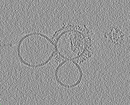 emd_8088.png
 http://ftp.pdbj.org/pub/emdb/structures/EMD-8088
 Sample components
 Processing
 Electron microscopy
