National Institutes of Health/National Institute of General Medical Sciences (NIH/NIGMS)
P41GM103832
United States
National Institutes of Health/National Institute Of Allergy and Infectious Diseases (NIH/NIAID)
AI057156
United States
National Institutes of Health/National Institute of General Medical Sciences (NIH/NIGMS)
R01GM080139
United States
Cancer Prevention and Research Institute of Texas (CPRIT)
RP140113
United States
Robert Welch
Q1242
United States
Citation
Journal: J Struct Biol / Year: 2016 Title: Alignment algorithms and per-particle CTF correction for single particle cryo-electron tomography. Authors: Jesús G Galaz-Montoya / Corey W Hecksel / Philip R Baldwin / Eryu Wang / Scott C Weaver / Michael F Schmid / Steven J Ludtke / Wah Chiu / Abstract: Single particle cryo-electron tomography (cryoSPT) extracts features from cryo-electron tomograms, followed by 3D classification, alignment and averaging to generate improved 3D density maps of such ...Single particle cryo-electron tomography (cryoSPT) extracts features from cryo-electron tomograms, followed by 3D classification, alignment and averaging to generate improved 3D density maps of such features. Robust methods to correct for the contrast transfer function (CTF) of the electron microscope are necessary for cryoSPT to reach its resolution potential. Many factors can make CTF correction for cryoSPT challenging, such as lack of eucentricity of the specimen stage, inherent low dose per image, specimen charging, beam-induced specimen motions, and defocus gradients resulting both from specimen tilting and from unpredictable ice thickness variations. Current CTF correction methods for cryoET make at least one of the following assumptions: that the defocus at the center of the image is the same across the images of a tiltseries, that the particles all lie at the same Z-height in the embedding ice, and/or that the specimen, the cryo-electron microscopy (cryoEM) grid and/or the carbon support are flat. These experimental conditions are not always met. We have developed a CTF correction algorithm for cryoSPT without making any of the aforementioned assumptions. We also introduce speed and accuracy improvements and a higher degree of automation to the subtomogram averaging algorithms available in EMAN2. Using motion-corrected images of isolated virus particles as a benchmark specimen, recorded with a DE20 direct detection camera, we show that our CTF correction and subtomogram alignment routines can yield subtomogram averages close to 4/5 Nyquist frequency of the detector under our experimental conditions.
History
Deposition
Feb 7, 2016
-
Header (metadata) release
Apr 13, 2016
-
Map release
Apr 13, 2016
-
Update
Jan 29, 2020
-
Current status
Jan 29, 2020
Processing site: RCSB / Status: Released
-
Structure visualization
Movie
Surface view with section colored by density value
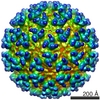
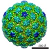
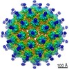
Shell ID: 1 / Name: Capsid / Diameter: 700.0 Å / T number (triangulation number): 4
-
Experimental details
-
Structure determination
Method
cryo EM
Processing
subtomogram averaging
Aggregation state
particle
-
Sample preparation
Buffer
pH: 7.4
Grid
Model: Quantifoil R1.2/1.3 / Material: COPPER / Mesh: 200 / Support film - #0 - Film type ID: 1 / Support film - #0 - Material: CARBON / Support film - #0 - topology: HOLEY / Support film - #1 - Film type ID: 2 / Support film - #1 - Material: CARBON / Support film - #1 - topology: CONTINUOUS / Pretreatment - Type: GLOW DISCHARGE
Film or detector model: DIRECT ELECTRON DE-20 (5k x 3k) / Detector mode: INTEGRATING / Average exposure time: 0.5 sec. / Average electron dose: 3.0 e/Å2
-
Image processing
Extraction
Number tomograms: 6 / Number images used: 516 / Method: Manual picking / Software - Name: EMAN2 (ver. 2.1)
In the structure databanks used in Yorodumi, some data are registered as the other names, "COVID-19 virus" and "2019-nCoV". Here are the details of the virus and the list of structure data.
Jan 31, 2019. EMDB accession codes are about to change! (news from PDBe EMDB page)
EMDB accession codes are about to change! (news from PDBe EMDB page)
The allocation of 4 digits for EMDB accession codes will soon come to an end. Whilst these codes will remain in use, new EMDB accession codes will include an additional digit and will expand incrementally as the available range of codes is exhausted. The current 4-digit format prefixed with “EMD-” (i.e. EMD-XXXX) will advance to a 5-digit format (i.e. EMD-XXXXX), and so on. It is currently estimated that the 4-digit codes will be depleted around Spring 2019, at which point the 5-digit format will come into force.
The EM Navigator/Yorodumi systems omit the EMD- prefix.
Related info.:Q: What is EMD? / ID/Accession-code notation in Yorodumi/EM Navigator
Yorodumi is a browser for structure data from EMDB, PDB, SASBDB, etc.
This page is also the successor to EM Navigator detail page, and also detail information page/front-end page for Omokage search.
The word "yorodu" (or yorozu) is an old Japanese word meaning "ten thousand". "mi" (miru) is to see.
Related info.:EMDB / PDB / SASBDB / Comparison of 3 databanks / Yorodumi Search / Aug 31, 2016. New EM Navigator & Yorodumi / Yorodumi Papers / Jmol/JSmol / Function and homology information / Changes in new EM Navigator and Yorodumi
 Movie
Movie Controller

 Yorodumi
 Open data
 Basic information
 Map data
 Sample
 Venezuelan equine encephalitis virus
 Authors
 United States, 5 items
  Citation
 Structure visualization
 Movie viewer
 Downloads & links
 emd_8071.png
 http://ftp.pdbj.org/pub/emdb/structures/EMD-8071
 Sample components
 Processing
 Electron microscopy
