 Movie
Movie Controller

[English] 日本語
 Yorodumi
- EMDB-8068: HKU1 spike with attached foldon domain and wild-type furin-cleava... -
+ Open data

- Basic information
| Entry | Database: EMDB / ID: EMD-8068 | |||||||||
|---|---|---|---|---|---|---|---|---|---|---|
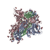
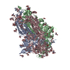
| Title | HKU1 spike with attached foldon domain and wild-type furin-cleavage site | |||||||||
 Map data Map data | None | |||||||||
 Sample Sample |
| |||||||||
| Biological species |  Human coronavirus HKU1 (isolate N5) Human coronavirus HKU1 (isolate N5) | |||||||||
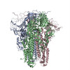
| Method | single particle reconstruction / negative staining / Resolution: 20.0 Å | |||||||||
 Authors Authors | Kirchdoerfer RN / Cottrell CA / Wang N / Pallesen J / Yassine HM / Turner HL / Corbett KS / Graham BS / McLellan JS / Ward AB | |||||||||
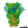
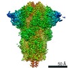
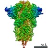
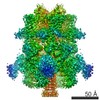
 Citation Citation | Journal: Nature / Year: 2016 Title: Pre-fusion structure of a human coronavirus spike protein. Authors: Robert N Kirchdoerfer / Christopher A Cottrell / Nianshuang Wang / Jesper Pallesen / Hadi M Yassine / Hannah L Turner / Kizzmekia S Corbett / Barney S Graham / Jason S McLellan / Andrew B Ward /  Abstract: HKU1 is a human betacoronavirus that causes mild yet prevalent respiratory disease, and is related to the zoonotic SARS and MERS betacoronaviruses, which have high fatality rates and pandemic ...HKU1 is a human betacoronavirus that causes mild yet prevalent respiratory disease, and is related to the zoonotic SARS and MERS betacoronaviruses, which have high fatality rates and pandemic potential. Cell tropism and host range is determined in part by the coronavirus spike (S) protein, which binds cellular receptors and mediates membrane fusion. As the largest known class I fusion protein, its size and extensive glycosylation have hindered structural studies of the full ectodomain, thus preventing a molecular understanding of its function and limiting development of effective interventions. Here we present the 4.0 Å resolution structure of the trimeric HKU1 S protein determined using single-particle cryo-electron microscopy. In the pre-fusion conformation, the receptor-binding subunits, S1, rest above the fusion-mediating subunits, S2, preventing their conformational rearrangement. Surprisingly, the S1 C-terminal domains are interdigitated and form extensive quaternary interactions that occlude surfaces known in other coronaviruses to bind protein receptors. These features, along with the location of the two protease sites known to be important for coronavirus entry, provide a structural basis to support a model of membrane fusion mediated by progressive S protein destabilization through receptor binding and proteolytic cleavage. These studies should also serve as a foundation for the structure-based design of betacoronavirus vaccine immunogens. | |||||||||
| History |
|
- Structure visualization
| Movie |
 Movie viewer Movie viewer |
|---|---|
| Structure viewer | EM map: SurfViewMolmilJmol/JSmol |
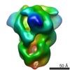
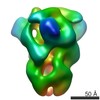
| Supplemental images |
 UCSF Chimera
- Downloads & links
-EMDB archive
| Map data | emd_8068.map.gz | 16.9 MB | EMDB map data format | |
|---|---|---|---|---|
| Header (meta data) | emd-8068-v30.xmlemd-8068.xml | 13.9 KB 13.9 KB | Display Display | EMDB header |
| Images | 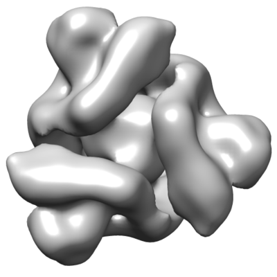 emd_8068.png emd_8068.png | 57.8 KB | ||
| Archive directory |  http://ftp.pdbj.org/pub/emdb/structures/EMD-8068ftp://ftp.pdbj.org/pub/emdb/structures/EMD-8068 http://ftp.pdbj.org/pub/emdb/structures/EMD-8068ftp://ftp.pdbj.org/pub/emdb/structures/EMD-8068 | HTTPS FTP |
-Validation report
| Summary document | emd_8068_validation.pdf.gz | 78.5 KB | Display | EMDB validaton report |
|---|---|---|---|---|
| Full document | emd_8068_full_validation.pdf.gz | 77.6 KB | Display | |
| Data in XML | emd_8068_validation.xml.gz | 493 B | Display | |
| Arichive directory | https://ftp.pdbj.org/pub/emdb/validation_reports/EMD-8068ftp://ftp.pdbj.org/pub/emdb/validation_reports/EMD-8068 | HTTPS FTP |
-Related structure data


-Links
| EMDB pages | EMDB (EBI/PDBe) / EMDataResource |
|---|
-Map
| File | Download / File: emd_8068.map.gz / Format: CCP4 / Size: 27 MB / Type: IMAGE STORED AS FLOATING POINT NUMBER (4 BYTES) | ||||||||||||||||||||||||||||||||||||||||||||||||||||||||||||
|---|---|---|---|---|---|---|---|---|---|---|---|---|---|---|---|---|---|---|---|---|---|---|---|---|---|---|---|---|---|---|---|---|---|---|---|---|---|---|---|---|---|---|---|---|---|---|---|---|---|---|---|---|---|---|---|---|---|---|---|---|---|
| Annotation | None | ||||||||||||||||||||||||||||||||||||||||||||||||||||||||||||
| Voxel size | X=Y=Z: 2.05 Å | ||||||||||||||||||||||||||||||||||||||||||||||||||||||||||||
| Density |
| ||||||||||||||||||||||||||||||||||||||||||||||||||||||||||||
| Symmetry | Space group: 1 | ||||||||||||||||||||||||||||||||||||||||||||||||||||||||||||
| Details | EMDB XML:
CCP4 map header:
| ||||||||||||||||||||||||||||||||||||||||||||||||||||||||||||
-Supplemental data
- Sample components
-Entire : HKU1 spike without foldon and with mutated furin-cleavage site
| Entire | Name: HKU1 spike without foldon and with mutated furin-cleavage site |
|---|---|
| Components |
|
-Supramolecule #1: HKU1 spike without foldon and with mutated furin-cleavage site
| Supramolecule | Name: HKU1 spike without foldon and with mutated furin-cleavage site type: complex / ID: 1 / Parent: 0 / Macromolecule list: all |
|---|---|
| Source (natural) | Organism: Human coronavirus HKU1 (isolate N5) |
| Recombinant expression | Organism:  Homo sapiens (human) / Recombinant cell: FreeStyle 293F / Recombinant plasmid: pVRC8400 Homo sapiens (human) / Recombinant cell: FreeStyle 293F / Recombinant plasmid: pVRC8400 |
| Molecular weight | Theoretical: 410 KDa |
-Macromolecule #1: HKU1 spike without foldon and with mutated furin-cleavage site
| Macromolecule | Name: HKU1 spike without foldon and with mutated furin-cleavage site type: protein_or_peptide / ID: 1 / Enantiomer: LEVO |
|---|---|
| Source (natural) | Organism: Human coronavirus HKU1 (isolate N5) |
| Recombinant expression | Organism: Homo sapiens (human) |
| Sequence | String: VIGDFNCTNS FINDYNKTIP RISEDVVDVS LGLGTYYVLN RVYLNTTLLF TGYFPKSGAN FRDLALKGSI YLSTLWYKPP FLSDFNNGIF SKVKNTKLYV NNTLYSEFST IVIGSVFVNT SYTIVVQPHN GILEITACQY TMCEYPHTVC KSKGSIRNES WHIDSSEPLC ...String: VIGDFNCTNS FINDYNKTIP RISEDVVDVS LGLGTYYVLN RVYLNTTLLF TGYFPKSGAN FRDLALKGSI YLSTLWYKPP FLSDFNNGIF SKVKNTKLYV NNTLYSEFST IVIGSVFVNT SYTIVVQPHN GILEITACQY TMCEYPHTVC KSKGSIRNES WHIDSSEPLC LFKKNFTYNV SADWLYFHFY QERGVFYAYY ADVGMPTTFL FSLYLGTILS HYYVMPLTCN AISSNTDNET LEYWVTPLSR RQYLLNFDEH GVITNAVDCS SSFLSEIQCK TQSFAPNTGV YDLSGFTVKP VATVYRRIPN LPDCDIDNWL NNVSVPSPLN WERRIFSNCN FNLSTLLRLV HVDSFSCNNL DKSKIFGSCF NSITVDKFAI PNRRRDDLQL GSSGFLQSSN YKIDISSSSC QLYYSLPLVN VTINNFNPSS WNRRYGFGSF NLSSYDVVYS DHCFSVNSDF CPCADPSVVN SCAKSKPPSA ICPAGTKYRH CDLDTTLYVK NWCRCSCLPD PISTYSPNTC PQKKVVVGIG EHCPGLGINE EKCGTQLNHS SCFCSPDAFL GWSFDSCISN NRCNIFSNFI FNGINSGTTC SNDLLYSNTE ISTGVCVNYD LYGITGQGIF KEVSAAYYNN WQNLLYDSNG NIIGFKDFLT NKTYTILPCY SGRVSAAFYQ NSSSPALLYR NLKCSYVLNN ISFISQPFYF DSYLGCVLNA VNLTSYSVSS CDLRMGSGFC IDYALPSSGG SGSGISSPYR FVTFEPFNVS FVNDSVETVG GLFEIQIPTN FTIAGHEEFI QTSSPKVTID CSAFVCSNYA ACHDLLSEYG TFCDNINSIL NEVNDLLDIT QLQVANALMQ GVTLSSNLNT NLHSDVDNID FKSLLGCLGS QCGSSSRSLL EDLLFNKVKL SDVGFVEAYN NCTGGSEIRD LLCVQSFNGI KVLPPILSET QISGYTTAAT VAAMFPPWSA AAGVPFSLNV QYRINGLGVT MDVLNKNQKL IANAFNKALL SIQNGFTATN SALAKIQSVV NANAQALNSL LQQLFNKFGA ISSSLQEILS RLDNLEAQVQ IDRLINGRLT ALNAYVSQQL SDITLIKAGA SRAIEKVNEC VKSQSPRINF CGNGNHILSL VQNAPYGLLF IHFSYKPTSF KTVLVSPGLC LSGDRGIAPK QGYFIKQNDS WMFTGSSYYY PEPISDKNVV FMNSCSVNFT KAPFIYLNNS IPNLSDFEAE LSLWFKNHTS IAPNLTSGRL EVLFQ |
-Experimental details
-Structure determination
| Method | negative staining |
|---|---|
 Processing Processing | single particle reconstruction |
| Aggregation state | particle |
-Sample preparation
| Concentration | 0.17 mg/mL | |||||||||
|---|---|---|---|---|---|---|---|---|---|---|
| Buffer | pH: 8 Component:
| |||||||||
| Staining | Type: NEGATIVE / Material: uranyl formate Details: 3uL sample was applied to grid for 30 seconds and then blotted. Grids were stained with 3uL 1% uranyl formate for 60 seconds followed by blotting. | |||||||||
| Grid | Material: COPPER / Mesh: 400 / Support film - Material: CARBON / Support film - topology: CONTINUOUS / Pretreatment - Type: GLOW DISCHARGE |
- Electron microscopy
| Microscope | FEI TECNAI SPIRIT |
|---|---|
| Image recording | Film or detector model: TVIPS TEMCAM-F416 (4k x 4k) / Number grids imaged: 1 / Number real images: 324 / Average exposure time: 1.0 sec. / Average electron dose: 28.0 e/Å2 Details: Images were collected using Legionon and processed using Appion. |
| Electron beam | Acceleration voltage: 120 kV / Electron source: LAB6 |
| Electron optics | Illumination mode: FLOOD BEAM / Imaging mode: BRIGHT FIELD / Nominal defocus max: 1.5 µm / Nominal defocus min: 1.0 µm / Nominal magnification: 52000 |
| Sample stage | Specimen holder model: SIDE ENTRY, EUCENTRIC |
| Experimental equipment |  Model: Tecnai Spirit / Image courtesy: FEI Company |
