 Movie
Movie Controller

[English] 日本語
 Yorodumi
- EMDB-8061: Structural basis of backwards motion in kinesin-14: plus-end dire... -
+ Open data

- Basic information
| Entry | Database: EMDB / ID: EMD-8061 | |||||||||
|---|---|---|---|---|---|---|---|---|---|---|
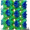
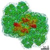
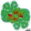
| Title | Structural basis of backwards motion in kinesin-14: plus-end directed nKn669 in the nucleotide-free state | |||||||||
 Map data Map data | None | |||||||||
 Sample Sample |
| |||||||||
 Keywords Keywords |  kinesin / kinesin-14 / microtubule / ATPase / STRUCTURAL PROTEIN-MOTOR PROTEIN complex kinesin / kinesin-14 / microtubule / ATPase / STRUCTURAL PROTEIN-MOTOR PROTEIN complex | |||||||||
| Function / homology |  Function and homology information Function and homology informationminus-end directed microtubule sliding / distributive segregation / regulation of mitotic spindle elongation / meiotic spindle assembly / mitotic spindle elongation / mitotic spindle microtubule / meiotic spindle organization / spindle assembly involved in female meiosis / minus-end-directed microtubule motor activity / regulation of mitotic spindle assembly ...minus-end directed microtubule sliding / distributive segregation / regulation of mitotic spindle elongation / meiotic spindle assembly / mitotic spindle elongation / mitotic spindle microtubule / meiotic spindle organization / spindle assembly involved in female meiosis / minus-end-directed microtubule motor activity / regulation of mitotic spindle assembly / microtubule bundle formation / meiotic spindle / mitotic centrosome separation / positive regulation of axon guidance / spindle organization / mitotic spindle assembly / mRNA transport / cytoplasmic microtubule / microtubule-based process / cellular response to interleukin-4 / mitotic spindle organization / chromosome segregation / Hydrolases; Acting on acid anhydrides; Acting on GTP to facilitate cellular and subcellular movement / Hydrolases; Acting on acid anhydrides; Acting on acid anhydrides to facilitate cellular and subcellular movement / structural constituent of cytoskeleton / spindle / microtubule cytoskeleton organization / microtubule cytoskeleton / double-stranded RNA binding / mitotic cell cycle / nervous system development / microtubule binding / microtubule / hydrolase activity / protein heterodimerization activity / cell division / GTPase activity / centrosome / ubiquitin protein ligase binding / GTP binding / protein homodimerization activity / ATP binding / metal ion binding / nucleus / cytosol / cytoplasmSimilarity search - Function | |||||||||
| Biological species |  Bos taurus (cattle) / Bos taurus (cattle) /  Drosophila melanogaster (fruit fly) Drosophila melanogaster (fruit fly) | |||||||||
| Method | helical reconstruction / cryo EM / Resolution: 5.8 Å | |||||||||
 Authors Authors | Shigematsu H / Yokoyama T / Kikkawa M / Shirouzu M / Nitta R | |||||||||
 Citation Citation | Journal: Structure / Year: 2016 Title: Structural Basis of Backwards Motion in Kinesin-1-Kinesin-14 Chimera: Implication for Kinesin-14 Motility. Authors: Masahiko Yamagishi / Hideki Shigematsu / Takeshi Yokoyama / Masahide Kikkawa / Mitsuhiro Sugawa / Mari Aoki / Mikako Shirouzu / Junichiro Yajima / Ryo Nitta /  Abstract: Kinesin-14 is a unique minus-end-directed microtubule-based motor. A swinging motion of a class-specific N-terminal neck helix has been proposed to produce minus-end directionality. However, it is ...Kinesin-14 is a unique minus-end-directed microtubule-based motor. A swinging motion of a class-specific N-terminal neck helix has been proposed to produce minus-end directionality. However, it is unclear how swinging of the neck helix is driven by ATP hydrolysis utilizing the highly conserved catalytic core among all kinesins. Here, using a motility assay, we show that in addition to the neck helix, the conserved five residues at the C-terminal region in kinesin-14, namely the neck mimic, are necessary to give kinesin-1 an ability to reverse its directionality toward the minus end of microtubules. Our structural analyses further demonstrate that the C-terminal neck mimic, in cooperation with conformational changes in the catalytic core during ATP binding, forms a kinesin-14 bundle with the N-terminal neck helix to swing toward the minus end of microtubules. Thus, the neck mimic plays a crucial role in coupling the chemical ATPase reaction with the mechanical cycle to produce the minus-end-directed motility of kinesin-14. | |||||||||
| History |
|
- Structure visualization
| Movie |
Movie viewer |
|---|---|
| Structure viewer | EM map: SurfViewMolmilJmol/JSmol |
| Supplemental images |

- Downloads & links
-EMDB archive
| Map data | emd_8061.map.gz | 10.7 MB | EMDB map data format | |
|---|---|---|---|---|
| Header (meta data) | emd-8061-v30.xmlemd-8061.xml | 14.9 KB 14.9 KB | Display Display | EMDB header |
| Images |  emd_8061.png emd_8061.png | 149.6 KB | ||
| Filedesc metadata | emd-8061.cif.gz | 6.8 KB | ||
| Archive directory |  http://ftp.pdbj.org/pub/emdb/structures/EMD-8061ftp://ftp.pdbj.org/pub/emdb/structures/EMD-8061 http://ftp.pdbj.org/pub/emdb/structures/EMD-8061ftp://ftp.pdbj.org/pub/emdb/structures/EMD-8061 | HTTPS FTP |
-Related structure data
| Related structure data |  5hnzMC  8058C  8059C 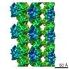 8060C  5hnwC  5hnxC  5hnyC M: atomic model generated by this map C: citing same article ( |
|---|---|
| Similar structure data |


-Links
| EMDB pages | EMDB (EBI/PDBe) / EMDataResource |
|---|---|
| Related items in Molecule of the Month |


-Map
| File | Download / File: emd_8061.map.gz / Format: CCP4 / Size: 12 MB / Type: IMAGE STORED AS FLOATING POINT NUMBER (4 BYTES) | ||||||||||||||||||||||||||||||||||||||||||||||||||||||||||||||||||||
|---|---|---|---|---|---|---|---|---|---|---|---|---|---|---|---|---|---|---|---|---|---|---|---|---|---|---|---|---|---|---|---|---|---|---|---|---|---|---|---|---|---|---|---|---|---|---|---|---|---|---|---|---|---|---|---|---|---|---|---|---|---|---|---|---|---|---|---|---|---|
| Annotation | None | ||||||||||||||||||||||||||||||||||||||||||||||||||||||||||||||||||||
| Voxel size | X=Y=Z: 1.284 Å | ||||||||||||||||||||||||||||||||||||||||||||||||||||||||||||||||||||
| Density |
| ||||||||||||||||||||||||||||||||||||||||||||||||||||||||||||||||||||
| Symmetry | Space group: 1 | ||||||||||||||||||||||||||||||||||||||||||||||||||||||||||||||||||||
| Details | EMDB XML:
CCP4 map header:
| ||||||||||||||||||||||||||||||||||||||||||||||||||||||||||||||||||||
-Supplemental data
- Sample components
+Entire : Plus-end directed Ncd chimera nKn669 in the nucleotide-free state...
+Supramolecule #1: Plus-end directed Ncd chimera nKn669 in the nucleotide-free state...
+Supramolecule #2: Tubulin alpha-1B chain
+Supramolecule #3: Tubulin beta-2B chain
+Supramolecule #4: Protein claret segregational,Plus-end directed kinesin-1/kinesin-...
+Macromolecule #1: Tubulin alpha-1B chain
+Macromolecule #2: Tubulin beta-2B chain
+Macromolecule #3: Protein claret segregational,Protein claret segregational,Plus-en...

+Macromolecule #4: MAGNESIUM ION
+Macromolecule #5: GUANOSINE-5'-TRIPHOSPHATE

+Macromolecule #6: GUANOSINE-5'-DIPHOSPHATE

+Macromolecule #7: TAXOL

-Experimental details
-Structure determination
| Method | cryo EM |
|---|---|
 Processing Processing | helical reconstruction |
| Aggregation state | filament |
-Sample preparation
| Buffer | pH: 6.8 |
|---|---|
| Vitrification | Cryogen name: ETHANE |
- Electron microscopy
| Microscope | FEI TECNAI ARCTICA |
|---|---|
| Electron beam | Acceleration voltage: 200 kV / Electron source: FIELD EMISSION GUN |
| Electron optics | Illumination mode: FLOOD BEAM / Imaging mode: BRIGHT FIELDBright-field microscopy |
| Image recording | Film or detector model: FEI FALCON II (4k x 4k) / Average electron dose: 30.0 e/Å2 |
| Experimental equipment |  Model: Talos Arctica / Image courtesy: FEI Company |
-Image processing
| Startup model | Type of model: EMDB MAP |
|---|---|
| Final angle assignment | Type: NOT APPLICABLE |
| Final reconstruction | Applied symmetry - Helical parameters - Δz: 8.77999 Å Applied symmetry - Helical parameters - Δ&Phi: -25.718312 ° Applied symmetry - Helical parameters - Axial symmetry: C1 (asymmetric) Resolution.type: BY AUTHOR / Resolution: 5.8 Å / Resolution method: FSC 0.143 CUT-OFF / Details: High-resolution noise substitution was performed / Number images used: 203826 |
-Atomic model buiding 1
| Refinement | Protocol: FLEXIBLE FIT |
|---|---|
| Output model | PDB-5hnz: |
