 Movie
Movie Controller


+ Open data

- Basic information
| Entry | Database: EMDB / ID: EMD-6473 | |||||||||
|---|---|---|---|---|---|---|---|---|---|---|
| Title | Platelet from a woman diagnosed with ovarian cancer | |||||||||
 Map data Map data | Tomographic reconstruction of a platelet from a woman with an ovarian malignancy | |||||||||
 Sample Sample |
| |||||||||
 Keywords Keywords | ovarian cancer / platelet / thrombosis / ultrastructure | |||||||||
| Biological species |  Homo sapiens (human) Homo sapiens (human) | |||||||||
| Method | electron tomography / cryo EM | |||||||||
 Authors Authors | Wang R / Stone RL / Kaelber JT / Rochat RH / Nick AM / Vijayan KV / Afshar-Kharghan V / Schmid MF / Dong JF / Sood AK / Chiu W | |||||||||
 Citation Citation | Journal: Proc Natl Acad Sci U S A / Year: 2015 Title: Electron cryotomography reveals ultrastructure alterations in platelets from patients with ovarian cancer. Authors: Rui Wang / Rebecca L Stone / Jason T Kaelber / Ryan H Rochat / Alpa M Nick / K Vinod Vijayan / Vahid Afshar-Kharghan / Michael F Schmid / Jing-Fei Dong / Anil K Sood / Wah Chiu /  Abstract: Thrombocytosis and platelet hyperreactivity are known to be associated with malignancy; however, there have been no ultrastructure studies of platelets from patients with ovarian cancer. Here, we ...Thrombocytosis and platelet hyperreactivity are known to be associated with malignancy; however, there have been no ultrastructure studies of platelets from patients with ovarian cancer. Here, we used electron cryotomography (cryo-ET) to examine frozen-hydrated platelets from patients with invasive ovarian cancer (n = 12) and control subjects either with benign adnexal mass (n = 5) or free from disease (n = 6). Qualitative inspections of the tomograms indicate significant morphological differences between the cancer and control platelets, including disruption of the microtubule marginal band. Quantitative analysis of subcellular features in 120 platelet electron tomograms from these two groups showed statistically significant differences in mitochondria, as well as microtubules. These structural variations in the platelets from the patients with cancer may be correlated with the altered platelet functions associated with malignancy. Cryo-ET of platelets shows potential as a noninvasive biomarker technology for ovarian cancer and other platelet-related diseases. | |||||||||
| History |
|
- Structure visualization
| Movie |
 Movie viewer Movie viewer |
|---|---|
| Supplemental images |

- Downloads & links
-EMDB archive
| Map data | emd_6473.map.gz | 13.5 GB | EMDB map data format | |
|---|---|---|---|---|
| Header (meta data) | emd-6473-v30.xmlemd-6473.xml | 9.2 KB 9.2 KB | Display Display | EMDB header |
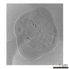
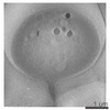
| Images | 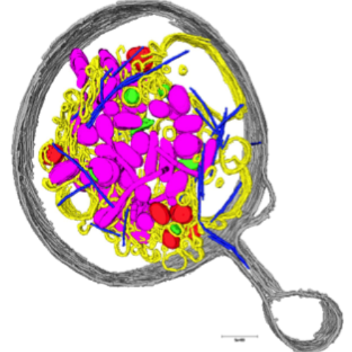 emd_6473.png emd_6473.png | 267.9 KB | ||
| Archive directory |  http://ftp.pdbj.org/pub/emdb/structures/EMD-6473ftp://ftp.pdbj.org/pub/emdb/structures/EMD-6473 http://ftp.pdbj.org/pub/emdb/structures/EMD-6473ftp://ftp.pdbj.org/pub/emdb/structures/EMD-6473 | HTTPS FTP |
-Related structure data
-Links
| EMDB pages | EMDB (EBI/PDBe) / EMDataResource |
|---|
-Map
| File | Download / File: emd_6473.map.gz / Format: CCP4 / Size: 14.8 GB / Type: IMAGE STORED AS SIGNED BYTE | ||||||||||||||||||||||||||||||||||||||||||||||||||||||||||||||||||||
|---|---|---|---|---|---|---|---|---|---|---|---|---|---|---|---|---|---|---|---|---|---|---|---|---|---|---|---|---|---|---|---|---|---|---|---|---|---|---|---|---|---|---|---|---|---|---|---|---|---|---|---|---|---|---|---|---|---|---|---|---|---|---|---|---|---|---|---|---|---|
| Annotation | Tomographic reconstruction of a platelet from a woman with an ovarian malignancy | ||||||||||||||||||||||||||||||||||||||||||||||||||||||||||||||||||||
| Voxel size | X=Y=Z: 14.6 Å | ||||||||||||||||||||||||||||||||||||||||||||||||||||||||||||||||||||
| Density |
| ||||||||||||||||||||||||||||||||||||||||||||||||||||||||||||||||||||
| Symmetry | Space group: 1 | ||||||||||||||||||||||||||||||||||||||||||||||||||||||||||||||||||||
| Details | EMDB XML:
CCP4 map header:
| ||||||||||||||||||||||||||||||||||||||||||||||||||||||||||||||||||||
-Supplemental data
- Sample components
-Entire : Tomographic reconstruction of a platelet from a woman with an ova...
| Entire | Name: Tomographic reconstruction of a platelet from a woman with an ovarian malignancy |
|---|---|
| Components |
|
-Supramolecule #1000: Tomographic reconstruction of a platelet from a woman with an ova...
| Supramolecule | Name: Tomographic reconstruction of a platelet from a woman with an ovarian malignancy type: sample / ID: 1000 / Number unique components: 1 |
|---|
-Supramolecule #1: platelet
| Supramolecule | Name: platelet / type: organelle_or_cellular_component / ID: 1 / Name.synonym: thrombocyte / Number of copies: 1 / Recombinant expression: No / Database: NCBI |
|---|---|
| Source (natural) | Organism: Homo sapiens (human) / synonym: human / Tissue: blood / Cell: platelet |
-Experimental details
-Structure determination
| Method | cryo EM |
|---|---|
 Processing Processing | electron tomography |
| Aggregation state | cell |
-Sample preparation
| Buffer | Details: native platelet-rich plasma |
|---|---|
| Grid | Details: 200 mesh Quantifoil R3.5/1 |
| Vitrification | Cryogen name: ETHANE / Chamber humidity: 100 % / Instrument: FEI VITROBOT MARK IV / Method: 1 blot, 3 seconds |
- Electron microscopy
| Microscope | JEOL 2200FSC |
|---|---|
| Electron beam | Acceleration voltage: 200 kV / Electron source: FIELD EMISSION GUN |
| Electron optics | Calibrated magnification: 10275 / Illumination mode: FLOOD BEAM / Imaging mode: BRIGHT FIELDBright-field microscopy / Cs: 2.0 mm / Nominal defocus max: 15.0 µm / Nominal magnification: 8000 |
| Specialist optics | Energy filter - Name: omega filter / Energy filter - Lower energy threshold: 0.0 eV / Energy filter - Upper energy threshold: 15.0 eV |
| Sample stage | Specimen holder model: GATAN LIQUID NITROGEN / Tilt series - Axis1 - Min angle: -62 ° / Tilt series - Axis1 - Max angle: 62 ° / Tilt series - Axis1 - Angle increment: 2 ° |
| Alignment procedure | Legacy - Astigmatism: objective lens astigmatism correction |
| Details | 2 degree tilt increment |
| Date | Feb 10, 2012 |
| Image recording | Category: CCD / Film or detector model: GATAN ULTRASCAN 4000 (4k x 4k) / Number real images: 62 / Average electron dose: 2 e/Å2 |
-Image processing
| Final reconstruction | Algorithm: OTHER / Software - Name: IMOD / Number images used: 62 |
|---|
