 Movie
Movie Controller

[English] 日本語
 Yorodumi
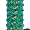
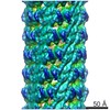
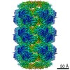
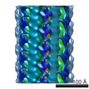
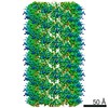
- EMDB-6325: 3D reconstruction of TMV at 4.5A from film micrographs using Frealix -
+ Open data

- Basic information
| Entry | Database: EMDB / ID: EMD-6325 | |||||||||
|---|---|---|---|---|---|---|---|---|---|---|
| Title | 3D reconstruction of TMV at 4.5A from film micrographs using Frealix | |||||||||
 Map data Map data | TMV map filtered using a negative B-factor, with amplitude matching against density derived from fitted atomic coordinates, a figure-of-merit filter, and subsequent helical symmetrization, as detailed in the primary citation. | |||||||||
 Sample Sample |
| |||||||||
| Biological species |    Tobacco mosaic virus Tobacco mosaic virus | |||||||||
| Method | helical reconstruction / cryo EM / Resolution: 4.5 Å | |||||||||
 Authors Authors | Rohou A / Grigorieff N | |||||||||
 Citation Citation | Journal: J Mol Biol / Year: 2007 Title: High-resolution electron microscopy of helical specimens: a fresh look at tobacco mosaic virus. Authors: Carsten Sachse / James Z Chen / Pierre-Damien Coureux / M Elizabeth Stroupe / Marcus Fändrich / Nikolaus Grigorieff /  Abstract: The treatment of helical objects as a string of single particles has become an established technique to resolve their three-dimensional (3D) structure using electron cryo-microscopy. It can be ...The treatment of helical objects as a string of single particles has become an established technique to resolve their three-dimensional (3D) structure using electron cryo-microscopy. It can be applied to a wide range of helical particles such as viruses, microtubules and helical filaments. We have made improvements to this approach using Tobacco Mosaic Virus (TMV) as a test specimen and obtained a map from 210,000 asymmetric units at a resolution better than 5 A. This was made possible by performing a full correction of the contrast transfer function of the microscope. Alignment of helical segments was helped by constraints derived from the helical symmetry of the virus. Furthermore, symmetrization was implemented by multiple inclusions of symmetry-related views in the 3D reconstruction. We used the density map to build an atomic model of TMV. The model was refined using a real-space refinement strategy that accommodates multiple conformers. The atomic model shows significant deviations from the deposited model for the helical form of TMV at the lower-radius region (residues 88 to 109). This region appears more ordered with well-defined secondary structure, compared with the earlier helical structure. The RNA phosphate backbone is sandwiched between two arginine side-chains, stabilizing the interaction between RNA and coat protein. A cluster of two or three carboxylates is buried in a hydrophobic environment isolating it from neighboring subunits. These carboxylates may represent the so-called Caspar carboxylates that form a metastable switch for viral disassembly. Overall, the observed differences suggest that the new model represents a different, more stable state of the virus, compared with the earlier published model. | |||||||||
| History |
|
- Structure visualization
| Movie |
 Movie viewer Movie viewer |
|---|---|
| Structure viewer | EM map: SurfViewMolmilJmol/JSmol |
| Supplemental images |

- Downloads & links
-EMDB archive
| Map data | emd_6325.map.gz | 28.3 MB | EMDB map data format | |
|---|---|---|---|---|
| Header (meta data) | emd-6325-v30.xmlemd-6325.xml | 10.4 KB 10.4 KB | Display Display | EMDB header |
| FSC (resolution estimation) | emd_6325_fsc.xml | 21.6 KB | Display | FSC data file |
| Images | 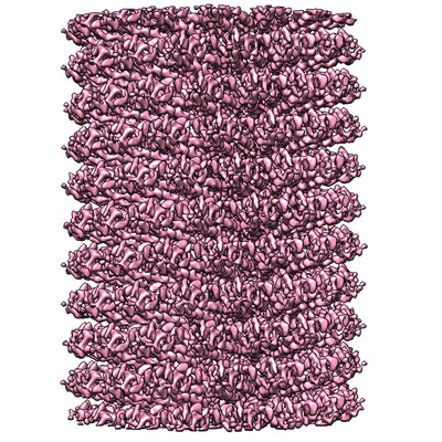 400_6325.gif 400_6325.gif 80_6325.gif 80_6325.gif | 130.1 KB 5.9 KB | ||
| Others | 3drec.map.gz | 398.2 MB | ||
| Archive directory |  http://ftp.pdbj.org/pub/emdb/structures/EMD-6325ftp://ftp.pdbj.org/pub/emdb/structures/EMD-6325 http://ftp.pdbj.org/pub/emdb/structures/EMD-6325ftp://ftp.pdbj.org/pub/emdb/structures/EMD-6325 | HTTPS FTP |
-Related structure data

-Links
| EMDB pages | EMDB (EBI/PDBe) / EMDataResource |
|---|---|
| Related items in Molecule of the Month |

-Map
| File | Download / File: emd_6325.map.gz / Format: CCP4 / Size: 29.8 MB / Type: IMAGE STORED AS FLOATING POINT NUMBER (4 BYTES) | ||||||||||||||||||||||||||||||||||||||||||||||||||||||||||||
|---|---|---|---|---|---|---|---|---|---|---|---|---|---|---|---|---|---|---|---|---|---|---|---|---|---|---|---|---|---|---|---|---|---|---|---|---|---|---|---|---|---|---|---|---|---|---|---|---|---|---|---|---|---|---|---|---|---|---|---|---|---|
| Annotation | TMV map filtered using a negative B-factor, with amplitude matching against density derived from fitted atomic coordinates, a figure-of-merit filter, and subsequent helical symmetrization, as detailed in the primary citation. | ||||||||||||||||||||||||||||||||||||||||||||||||||||||||||||
| Voxel size | X=Y=Z: 1.163 Å | ||||||||||||||||||||||||||||||||||||||||||||||||||||||||||||
| Density |
| ||||||||||||||||||||||||||||||||||||||||||||||||||||||||||||
| Symmetry | Space group: 1 | ||||||||||||||||||||||||||||||||||||||||||||||||||||||||||||
| Details | EMDB XML:
CCP4 map header:
| ||||||||||||||||||||||||||||||||||||||||||||||||||||||||||||
-Supplemental data
-Supplemental map: 3drec.map
| File | 3drec.map | ||||||||||||
|---|---|---|---|---|---|---|---|---|---|---|---|---|---|
| Projections & Slices |
| ||||||||||||
| Density Histograms |
 Z
Z Y
Y X
X


- Sample components
-Entire : Tobacco mosaic virus
| Entire | Name: Tobacco mosaic virus |
|---|---|
| Components |
|
-Supramolecule #1000: Tobacco mosaic virus
| Supramolecule | Name: Tobacco mosaic virus / type: sample / ID: 1000 / Oligomeric state: Helical assembly / Number unique components: 1 |
|---|
-Supramolecule #1: Tobacco mosaic virus
| Supramolecule | Name: Tobacco mosaic virus / type: virus / ID: 1 / NCBI-ID: 12242 / Sci species name: Tobacco mosaic virus / Sci species strain: vulgare / Database: NCBI / Virus type: VIRION / Virus isolate: STRAIN / Virus enveloped: No / Virus empty: No |
|---|---|
| Host (natural) | Organism:  Nicotiana tabacum (common tobacco) / synonym: PLANTAE(HIGHER PLANTS) Nicotiana tabacum (common tobacco) / synonym: PLANTAE(HIGHER PLANTS) |
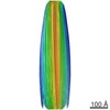
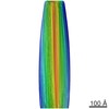
| Virus shell | Shell ID: 1 / Name: Virus shell 1 / Diameter: 180 Å |
-Experimental details
-Structure determination
| Method | cryo EM |
|---|---|
 Processing Processing | helical reconstruction |
| Aggregation state | filament |
-Sample preparation
| Concentration | 2.5 mg/mL |
|---|---|
| Buffer | Details: Phosphate, 5 mM EDTA |
| Grid | Details: Quantifoil grid |
| Vitrification | Cryogen name: ETHANE / Instrument: HOMEMADE PLUNGER / Method: Blot for 2 seconds before plunging |
- Electron microscopy
| Microscope | FEI TECNAI F30 |
|---|---|
| Electron beam | Acceleration voltage: 200 kV / Electron source: FIELD EMISSION GUN |
| Electron optics | Illumination mode: FLOOD BEAM / Imaging mode: BRIGHT FIELDBright-field microscopy / Cs: 2.0 mm / Nominal defocus max: 3.86 µm / Nominal defocus min: 1.688 µm / Nominal magnification: 59000 |
| Sample stage | Specimen holder model: GATAN LIQUID NITROGEN |
| Date | Nov 22, 2005 |
| Image recording | Category: FILM / Film or detector model: KODAK SO-163 FILM / Digitization - Scanner: ZEISS SCAI / Digitization - Sampling interval: 7.0 µm / Number real images: 7 / Average electron dose: 15 e/Å2 |
| Experimental equipment |  Model: Tecnai F30 / Image courtesy: FEI Company |
-Image processing
| CTF correction | Details: Defocus estimated for each helical subunit |
|---|---|
| Final reconstruction | Applied symmetry - Helical parameters - Δz: 1.39259 Å Applied symmetry - Helical parameters - Δ&Phi: 22.0318 ° Applied symmetry - Helical parameters - Axial symmetry: C1 (asymmetric) Algorithm: OTHER / Resolution.type: BY AUTHOR / Resolution: 4.5 Å / Resolution method: OTHER / Software - Name: Frealix Details: Only data up to 5.5 Angstrom were used during refinement in the final rounds. In earlier rounds, this threshold was set to a lower resolution. |
| Details | Filaments were processed using Frealix assuming constant helical parameters and without continuity restraints on the helical parameters. |
| FSC plot (resolution estimation) |  |
