 Movie
Movie Controller


+ Open data

- Basic information
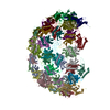
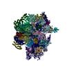
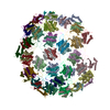
| Entry | Database: EMDB / ID: EMD-6307 | |||||||||
|---|---|---|---|---|---|---|---|---|---|---|
| Title | Electron cryo-microscopy of microtubule-bound TTLL7 | |||||||||
 Map data Map data | Reconstruction of a Tubulin Tyrosine Ligase-Like Glutamylase bound to the microtubule | |||||||||
 Sample Sample |
| |||||||||
 Keywords Keywords | microtubule-bound TTLL7 | |||||||||
| Biological species |   Homo sapiens (human) / Homo sapiens (human) /  Bos taurus (cattle) Bos taurus (cattle) | |||||||||
| Method | helical reconstruction / cryo EM / Resolution: 7.95 Å | |||||||||
 Authors Authors | Wilson-Kubalek EM / Garnham CP / Vemu A / Yu I / Szyk A / Lander GC / Milligan RA / Roll-Mecak A | |||||||||
 Citation Citation | Journal: Cell / Year: 2015 Title: Multivalent Microtubule Recognition by Tubulin Tyrosine Ligase-like Family Glutamylases. Authors: Christopher P Garnham / Annapurna Vemu / Elizabeth M Wilson-Kubalek / Ian Yu / Agnieszka Szyk / Gabriel C Lander / Ronald A Milligan / Antonina Roll-Mecak /  Abstract: Glutamylation, the most prevalent tubulin posttranslational modification, marks stable microtubules and regulates recruitment and activity of microtubule- interacting proteins. Nine enzymes of the ...Glutamylation, the most prevalent tubulin posttranslational modification, marks stable microtubules and regulates recruitment and activity of microtubule- interacting proteins. Nine enzymes of the tubulin tyrosine ligase-like (TTLL) family catalyze glutamylation. TTLL7, the most abundant neuronal glutamylase, adds glutamates preferentially to the β-tubulin tail. Coupled with ensemble and single-molecule biochemistry, our hybrid X-ray and cryo-electron microscopy structure of TTLL7 bound to the microtubule delineates a tripartite microtubule recognition strategy. The enzyme uses its core to engage the disordered anionic tails of α- and β-tubulin, and a flexible cationic domain to bind the microtubule and position itself for β-tail modification. Furthermore, we demonstrate that all single-chain TTLLs with known glutamylase activity utilize a cationic microtubule-binding domain analogous to that of TTLL7. Therefore, our work reveals the combined use of folded and intrinsically disordered substrate recognition elements as the molecular basis for specificity among the enzymes primarily responsible for chemically diversifying cellular microtubules. | |||||||||
| History |
|
- Structure visualization
| Movie |
 Movie viewer Movie viewer |
|---|---|
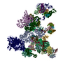
| Structure viewer | EM map: SurfViewMolmilJmol/JSmol |
| Supplemental images |

- Downloads & links
-EMDB archive
| Map data | emd_6307.map.gz | 1.5 MB | EMDB map data format | |
|---|---|---|---|---|
| Header (meta data) | emd-6307-v30.xmlemd-6307.xml | 10.7 KB 10.7 KB | Display Display | EMDB header |
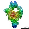
| Images | 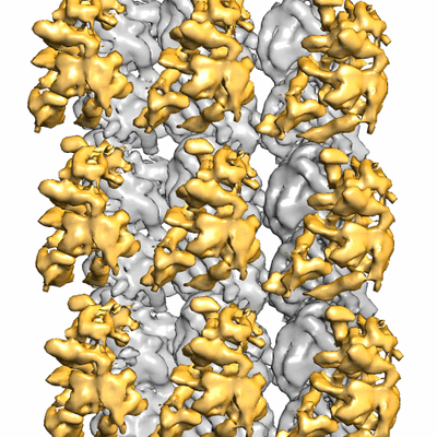 400_6307.gif 400_6307.gif 80_6307.gif 80_6307.gif | 123.6 KB 7.1 KB | ||
| Archive directory |  http://ftp.pdbj.org/pub/emdb/structures/EMD-6307ftp://ftp.pdbj.org/pub/emdb/structures/EMD-6307 http://ftp.pdbj.org/pub/emdb/structures/EMD-6307ftp://ftp.pdbj.org/pub/emdb/structures/EMD-6307 | HTTPS FTP |
-Related structure data


-Links
| EMDB pages | EMDB (EBI/PDBe) / EMDataResource |
|---|
-Map
| File | Download / File: emd_6307.map.gz / Format: CCP4 / Size: 1.6 MB / Type: IMAGE STORED AS FLOATING POINT NUMBER (4 BYTES) | ||||||||||||||||||||||||||||||||||||||||||||||||||||||||||||
|---|---|---|---|---|---|---|---|---|---|---|---|---|---|---|---|---|---|---|---|---|---|---|---|---|---|---|---|---|---|---|---|---|---|---|---|---|---|---|---|---|---|---|---|---|---|---|---|---|---|---|---|---|---|---|---|---|---|---|---|---|---|
| Annotation | Reconstruction of a Tubulin Tyrosine Ligase-Like Glutamylase bound to the microtubule | ||||||||||||||||||||||||||||||||||||||||||||||||||||||||||||
| Voxel size | X=Y=Z: 2.73 Å | ||||||||||||||||||||||||||||||||||||||||||||||||||||||||||||
| Density |
| ||||||||||||||||||||||||||||||||||||||||||||||||||||||||||||
| Symmetry | Space group: 1 | ||||||||||||||||||||||||||||||||||||||||||||||||||||||||||||
| Details | EMDB XML:
CCP4 map header:
| ||||||||||||||||||||||||||||||||||||||||||||||||||||||||||||
-Supplemental data
- Sample components
-Entire : Tubulin Tyrosine Ligase-Like (TTLL7) bound to the microtubule
| Entire | Name: Tubulin Tyrosine Ligase-Like (TTLL7) bound to the microtubule |
|---|---|
| Components |
|
-Supramolecule #1000: Tubulin Tyrosine Ligase-Like (TTLL7) bound to the microtubule
| Supramolecule | Name: Tubulin Tyrosine Ligase-Like (TTLL7) bound to the microtubule type: sample / ID: 1000 / Details: The sample was monodisperse / Oligomeric state: one TTLL7 bound to tubulin dimer / Number unique components: 2 |
|---|---|
| Molecular weight | Experimental: 61 KDa / Theoretical: 61 KDa / Method: SDS PAGE |
-Macromolecule #1: Tubulin Tyrosine Ligase-Like (TTLL7) Family Glutamylase
| Macromolecule | Name: Tubulin Tyrosine Ligase-Like (TTLL7) Family Glutamylase type: protein_or_peptide / ID: 1 / Name.synonym: TTLL7 / Number of copies: 1 / Oligomeric state: monomer / Recombinant expression: Yes |
|---|---|
| Source (natural) | Organism: Homo sapiens (human) / synonym: Human |
| Molecular weight | Experimental: 61 KDa / Theoretical: 61 KDa |
| Recombinant expression | Organism:  Escherichia coli (E. coli) Escherichia coli (E. coli) |
-Macromolecule #2: tubulin
| Macromolecule | Name: tubulin / type: protein_or_peptide / ID: 2 Details: Two sources of tubulin were used: cytoskeletal tubulin from bovine brain and human tubulin from TCA 201 cells. Recombinant expression: No / Database: NCBI |
|---|---|
| Source (natural) | Organism: Bos taurus (cattle) / synonym: bovine / Tissue: brain / Location in cell: cytoskeleton |
-Experimental details
-Structure determination
| Method | cryo EM |
|---|---|
 Processing Processing | helical reconstruction |
| Aggregation state | particle |
-Sample preparation
| Concentration | 11 mg/mL |
|---|---|
| Buffer | pH: 7 / Details: 20 mM HEPES, 0.5 mM ATP, 1 mM TCEP, 50 mM NaCl |
| Grid | Details: Protochips C-flat grid: holey carbon with 2 um holes and 400 mesh copper grid with 2 um spacing |
| Vitrification | Cryogen name: ETHANE / Chamber humidity: 80 % / Chamber temperature: 93 K / Instrument: HOMEMADE PLUNGER Method: Blot grid from behind for 3 seconds before plunging. |
- Electron microscopy
| Microscope | FEI TECNAI F20 |
|---|---|
| Electron beam | Acceleration voltage: 200 kV / Electron source: FIELD EMISSION GUN |
| Electron optics | Illumination mode: FLOOD BEAM / Imaging mode: BRIGHT FIELDBright-field microscopy / Nominal defocus max: 2.5 µm / Nominal defocus min: 0.5 µm / Nominal magnification: 62000 |
| Sample stage | Specimen holder model: GATAN LIQUID NITROGEN |
| Alignment procedure | Legacy - Astigmatism: Objective lens astigmatism was corrected at 62000x magnification. |
| Date | Aug 5, 2014 |
| Image recording | Category: CCD / Film or detector model: TVIPS TEMCAM-F416 (4k x 4k) / Digitization - Sampling interval: 2.73 µm / Number real images: 614 / Average electron dose: 20 e/Å2 / Bits/pixel: 16 |
| Experimental equipment |  Model: Tecnai F20 / Image courtesy: FEI Company |
-Image processing
| CTF correction | Details: CTFIND v3 |
|---|---|
| Final reconstruction | Algorithm: OTHER / Resolution.type: BY AUTHOR / Resolution: 7.95 Å / Resolution method: OTHER / Software - Name: EMAN2, FREALIGN Details: Final maps were calculated from three averaged data sets. |
| Details | We used IHRSR adapted for microtubules with a dimer repeat. |
