 Movie
Movie Controller

[English] 日本語
 Yorodumi
- EMDB-6289: Three dimensional reconstruction of bovine dynactin complex by cr... -
+ Open data

- Basic information
| Entry | Database: EMDB / ID: EMD-6289 | |||||||||
|---|---|---|---|---|---|---|---|---|---|---|
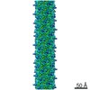
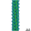
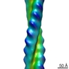
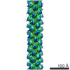
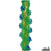
| Title | Three dimensional reconstruction of bovine dynactin complex by cryo electron microscopy | |||||||||
 Map data Map data | Three dimensional reconstruction of bovine dynactin complex by cryo electron microscopy | |||||||||
 Sample Sample |
| |||||||||
 Keywords Keywords |  dynactin / actin-related proteins / dynein dynactin / actin-related proteins / dynein | |||||||||
| Biological species |  Bos taurus (cattle) Bos taurus (cattle) | |||||||||
| Method | single particle reconstruction / cryo EM / Resolution: 6.5 Å | |||||||||
 Authors Authors | Chowdhury S / Ketcham SA / Schroer TA / Lander GC | |||||||||
 Citation Citation | Journal: Nat Struct Mol Biol / Year: 2015 Title: Structural organization of the dynein-dynactin complex bound to microtubules. Authors: Saikat Chowdhury / Stephanie A Ketcham / Trina A Schroer / Gabriel C Lander /  Abstract: Cytoplasmic dynein associates with dynactin to drive cargo movement on microtubules, but the structure of the dynein-dynactin complex is unknown. Using electron microscopy, we determined the ...Cytoplasmic dynein associates with dynactin to drive cargo movement on microtubules, but the structure of the dynein-dynactin complex is unknown. Using electron microscopy, we determined the organization of native bovine dynein, dynactin and the dynein-dynactin-microtubule quaternary complex. In the microtubule-bound complex, the dynein motor domains are positioned for processive unidirectional movement, and the cargo-binding domains of both dynein and dynactin are accessible. | |||||||||
| History |
|
- Structure visualization
| Movie |
 Movie viewer Movie viewer |
|---|---|
| Structure viewer | EM map: SurfViewMolmilJmol/JSmol |
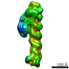
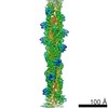
| Supplemental images |

- Downloads & links
-EMDB archive
| Map data | emd_6289.map.gz | 59.4 MB | EMDB map data format | |
|---|---|---|---|---|
| Header (meta data) | emd-6289-v30.xmlemd-6289.xml | 18.1 KB 18.1 KB | Display Display | EMDB header |
| Images | 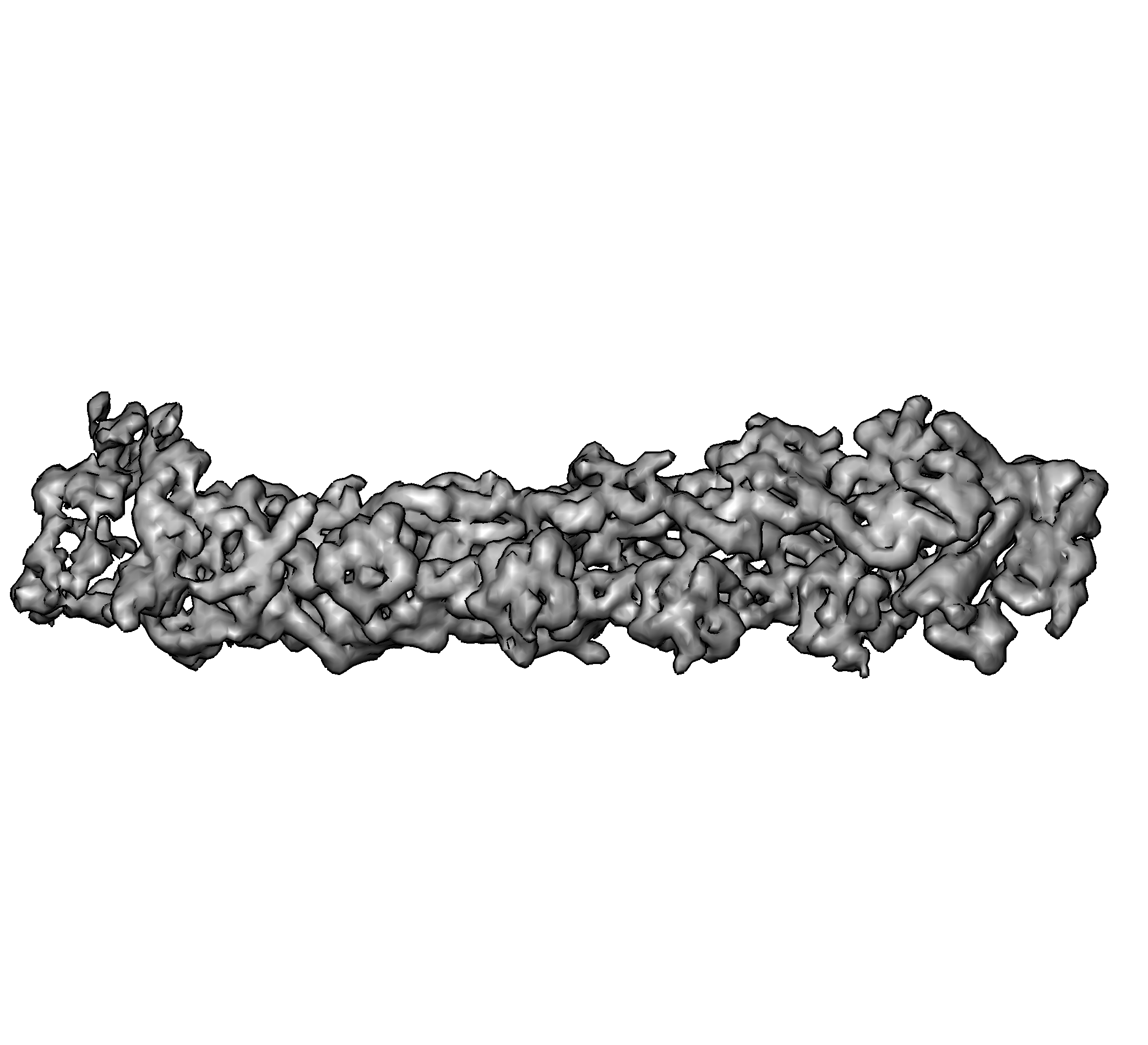 emd_6289.png emd_6289.png | 455 KB | ||
| Archive directory |  http://ftp.pdbj.org/pub/emdb/structures/EMD-6289ftp://ftp.pdbj.org/pub/emdb/structures/EMD-6289 http://ftp.pdbj.org/pub/emdb/structures/EMD-6289ftp://ftp.pdbj.org/pub/emdb/structures/EMD-6289 | HTTPS FTP |
-Related structure data
-Links
| EMDB pages | EMDB (EBI/PDBe) / EMDataResource |
|---|
-Map
| File | Download / File: emd_6289.map.gz / Format: CCP4 / Size: 64 MB / Type: IMAGE STORED AS FLOATING POINT NUMBER (4 BYTES) | ||||||||||||||||||||||||||||||||||||||||||||||||||||||||||||||||||||
|---|---|---|---|---|---|---|---|---|---|---|---|---|---|---|---|---|---|---|---|---|---|---|---|---|---|---|---|---|---|---|---|---|---|---|---|---|---|---|---|---|---|---|---|---|---|---|---|---|---|---|---|---|---|---|---|---|---|---|---|---|---|---|---|---|---|---|---|---|---|
| Annotation | Three dimensional reconstruction of bovine dynactin complex by cryo electron microscopy | ||||||||||||||||||||||||||||||||||||||||||||||||||||||||||||||||||||
| Voxel size | X=Y=Z: 2.62 Å | ||||||||||||||||||||||||||||||||||||||||||||||||||||||||||||||||||||
| Density |
| ||||||||||||||||||||||||||||||||||||||||||||||||||||||||||||||||||||
| Symmetry | Space group: 1 | ||||||||||||||||||||||||||||||||||||||||||||||||||||||||||||||||||||
| Details | EMDB XML:
CCP4 map header:
| ||||||||||||||||||||||||||||||||||||||||||||||||||||||||||||||||||||
-Supplemental data
- Sample components
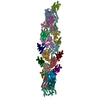
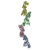
-Entire : Three-dimensional reconstruction of bovine dynactin complex by cryo EM
| Entire | Name: Three-dimensional reconstruction of bovine dynactin complex by cryo EM |
|---|---|
| Components |
|
-Supramolecule #1000: Three-dimensional reconstruction of bovine dynactin complex by cryo EM
| Supramolecule | Name: Three-dimensional reconstruction of bovine dynactin complex by cryo EM type: sample / ID: 1000 Details: The sample was monodisperse. The shoulder domain of dynactin would dissociate during cryo grid preparation. Oligomeric state: One CapZ alpha, one capZ beta, eight Arp1, one actin, one Arp11, one p25, one p27, one p62 Number unique components: 8 |
|---|---|
| Molecular weight | Theoretical: 1 MDa |
-Macromolecule #1: Arp1
| Macromolecule | Name: Arp1 / type: protein_or_peptide / ID: 1 / Number of copies: 8 / Oligomeric state: Octamer / Recombinant expression: No / Database: NCBI |
|---|---|
| Source (natural) | Organism: Bos taurus (cattle) / synonym: bovine / Tissue: Brain / Cell: Neuron / Location in cell: Cytoplasm |
| Molecular weight | Theoretical: 42.6 KDa |
-Macromolecule #2: Actin
| Macromolecule | Name: Actin / type: protein_or_peptide / ID: 2 / Number of copies: 1 / Oligomeric state: Monomer / Recombinant expression: No / Database: NCBI |
|---|---|
| Source (natural) | Organism: Bos taurus (cattle) / synonym: bovine / Tissue: Brain / Cell: Neuron / Location in cell: Cytoplasm |
| Molecular weight | Theoretical: 42 KDa |
-Macromolecule #3: Arp11
| Macromolecule | Name: Arp11 / type: protein_or_peptide / ID: 3 / Number of copies: 1 / Oligomeric state: Monomer / Recombinant expression: No / Database: NCBI |
|---|---|
| Source (natural) | Organism: Bos taurus (cattle) / synonym: bovine / Tissue: Brain / Cell: Neuron / Location in cell: Cytoplasm |
| Molecular weight | Theoretical: 42 KDa |
-Macromolecule #4: p62
| Macromolecule | Name: p62 / type: protein_or_peptide / ID: 4 / Number of copies: 1 / Oligomeric state: Monomer / Recombinant expression: No / Database: NCBI |
|---|---|
| Source (natural) | Organism: Bos taurus (cattle) / synonym: bovine / Tissue: Brain / Cell: Neuron / Location in cell: Cytoplasm |
| Molecular weight | Theoretical: 62 KDa |
-Macromolecule #5: p25
| Macromolecule | Name: p25 / type: protein_or_peptide / ID: 5 / Number of copies: 1 / Oligomeric state: Monomer / Recombinant expression: No / Database: NCBI |
|---|---|
| Source (natural) | Organism: Bos taurus (cattle) / synonym: bovine / Tissue: Brain / Cell: Neuron / Location in cell: Cytoplasm |
| Molecular weight | Theoretical: 25 KDa |
-Macromolecule #6: p27
| Macromolecule | Name: p27 / type: protein_or_peptide / ID: 6 / Number of copies: 1 / Oligomeric state: Monomer / Recombinant expression: No / Database: NCBI |
|---|---|
| Source (natural) | Organism: Bos taurus (cattle) / synonym: bovine / Tissue: Brain / Cell: Neuron / Location in cell: Cytoplasm |
| Molecular weight | Theoretical: 27 KDa |
-Macromolecule #7: CapZ alpha
| Macromolecule | Name: CapZ alpha / type: protein_or_peptide / ID: 7 / Number of copies: 1 / Oligomeric state: Monomer / Recombinant expression: No / Database: NCBI |
|---|---|
| Source (natural) | Organism: Bos taurus (cattle) / synonym: bovine / Tissue: Brain / Cell: Neuron / Location in cell: Cytoplasm |
| Molecular weight | Theoretical: 33 KDa |
-Macromolecule #8: CapZ beta
| Macromolecule | Name: CapZ beta / type: protein_or_peptide / ID: 8 / Number of copies: 1 / Oligomeric state: Monomer / Recombinant expression: No / Database: NCBI |
|---|---|
| Source (natural) | Organism: Bos taurus (cattle) / synonym: bovine / Tissue: Brain / Cell: Neuron / Location in cell: Cytoplasm |
| Molecular weight | Theoretical: 33 KDa |
-Experimental details
-Structure determination
| Method | cryo EM |
|---|---|
 Processing Processing | single particle reconstruction |
| Aggregation state | particle |
-Sample preparation
| Concentration | 0.05 mg/mL |
|---|---|
| Buffer | pH: 7.2 / Details: 35 mM Tris, 5 mM MgSO4, 150 mM KCl, 1 mM TCEP |
| Grid | Details: 400 mesh C-flat holey carbon grid |
| Vitrification | Cryogen name: ETHANE / Chamber humidity: 98 % / Chamber temperature: 83.15 K / Instrument: HOMEMADE PLUNGER Details: External environment at 4 degrees Celsius, 98% humidity. Method: 4 uL dynactin sample was applied to a cflat holey grid. Excess sample was manually blotted using filter paper for 5-7 seconds and immediately vitrified by plunge-freezing into liquid ethane ...Method: 4 uL dynactin sample was applied to a cflat holey grid. Excess sample was manually blotted using filter paper for 5-7 seconds and immediately vitrified by plunge-freezing into liquid ethane slurry at -179 degrees Celsius. |
- Electron microscopy
| Microscope | FEI TITAN KRIOS |
|---|---|
| Electron beam | Acceleration voltage: 300 kV / Electron source: FIELD EMISSION GUN |
| Electron optics | Calibrated magnification: 22500 / Illumination mode: FLOOD BEAM / Imaging mode: BRIGHT FIELDBright-field microscopy / Cs: 2.7 mm / Nominal defocus max: 4.0 µm / Nominal defocus min: 0.8 µm / Nominal magnification: 22500 |
| Sample stage | Specimen holder: Liquid nitrogen-cooled / Specimen holder model: FEI TITAN KRIOS AUTOGRID HOLDER |
| Temperature | Min: 83 K / Max: 85 K / Average: 84 K |
| Alignment procedure | Legacy - Astigmatism: Objective astigmatism was corrected using a quadrupole stigmator at 22,500 times magnification. |
| Details | Imaging was done on a Gatan K2 Summit camera operated in counting mode at a dose rate of 10 electrons/pixel/s. Each movie comprised 30 frames acquired over 6s, with a cumulative dose of 35 electrons/A**2. |
| Date | Oct 20, 2014 |
| Image recording | Category: CCD / Film or detector model: GATAN K2 (4k x 4k) / Number real images: 2421 / Average electron dose: 35 e/Å2 Details: Data collected using Leginon automated image acquisition software. Each movie comprised 30 frames acquired over 6 seconds. |
| Experimental equipment |  Model: Titan Krios / Image courtesy: FEI Company |
-Image processing
| CTF correction | Details: Each particle |
|---|---|
| Final angle assignment | Details: Theta 45 degrees, phi 45 degrees |
| Final reconstruction | Algorithm: OTHER / Resolution.type: BY AUTHOR / Resolution: 6.5 Å / Resolution method: OTHER / Software - Name: Relion Details: Processing leading up to 3D reconstruction was performed using the Appion package. Particles were selected from micrographs using a template-based automated particle picker. The stack was ...Details: Processing leading up to 3D reconstruction was performed using the Appion package. Particles were selected from micrographs using a template-based automated particle picker. The stack was subjected to five iterations of iterative 2D alignment and classification using multivariate statistical analysis (MSA) and multi-reference alignment (MRA). The clean particle stack was subjected to 25 iterations of 3D classification with three classes using the Relion suite. Particles belonging to well-resolved 3D class averages were used for further refinement by projection matching in Relion. Number images used: 59538 |
| Details | Processing leading up to 3D reconstruction was performed using the Appion package. Particles were selected from micrographs using a template-based automated particle picker. The stack was subjected to five iterations of iterative 2D alignment and classification using multivariate statistical analysis (MSA) and multi-reference alignment (MRA). The clean particle stack was subjected to 25 iterations of 3D classification with three classes using the Relion suite. Particles belonging to well-resolved 3D class averages were used for further refinement by projection matching in Relion. |
