- EMDB-5923: Architecture and assembly of the archaeal Cdc48-20S proteasome -
+
Open data
ID or keywords:
Loading...
-
Basic information
Entry
Database: EMDB / ID: EMD-5923
Title
Architecture and assembly of the archaeal Cdc48-20S proteasome
Map data
Single-particle reconstruction of the archaeal Cdc48-20S proteasome
Sample
Sample: Archaeal proteasome Cdc48-20S
Protein or peptide: Cdc48
Protein or peptide: 20S alpha subunit
Protein or peptide: 20S beta subunit
Keywords
Archaeal proteasome / Cdc48 / architecture / single-particle EM
Function / homology
Function and homology information
macromolecule metabolic process / primary metabolic process / response to stimulus / : / proteasome core complex, alpha-subunit complex / threonine-type endopeptidase activity / proteasomal protein catabolic process / ubiquitin-dependent protein catabolic process / endopeptidase activity / ATP hydrolysis activity ...macromolecule metabolic process / primary metabolic process / response to stimulus / : / proteasome core complex, alpha-subunit complex / threonine-type endopeptidase activity / proteasomal protein catabolic process / ubiquitin-dependent protein catabolic process / endopeptidase activity / ATP hydrolysis activity / ATP binding / cytoplasm Similarity search - Function
Proteasome alpha subunit, archaeal / AAA ATPase, CDC48 family / Cell division protein 48 (CDC48), N-terminal domain / CDC48, N-terminal subdomain / Cell division protein 48 (CDC48) N-terminal domain / CDC48, domain 2 / Cell division protein 48 (CDC48), domain 2 / Cell division protein 48 (CDC48) domain 2 / CDC48 domain 2-like superfamily / Aspartate decarboxylase-like domain superfamily ...Proteasome alpha subunit, archaeal / AAA ATPase, CDC48 family / Cell division protein 48 (CDC48), N-terminal domain / CDC48, N-terminal subdomain / Cell division protein 48 (CDC48) N-terminal domain / CDC48, domain 2 / Cell division protein 48 (CDC48), domain 2 / Cell division protein 48 (CDC48) domain 2 / CDC48 domain 2-like superfamily / Aspartate decarboxylase-like domain superfamily / Proteasome subunit A N-terminal signature / Proteasome alpha-type subunits signature. / Proteasome alpha-subunit, N-terminal domain / Proteasome subunit A N-terminal signature Add an annotation / Proteasome alpha-type subunit / Proteasome alpha-type subunit profile. / Proteasome subunit / Proteasome, subunit alpha/beta / AAA ATPase, AAA+ lid domain / AAA+ lid domain / ATPase, AAA-type, conserved site / AAA-protein family signature. / Nucleophile aminohydrolases, N-terminal / ATPase family associated with various cellular activities (AAA) / ATPase, AAA-type, core / ATPases associated with a variety of cellular activities / AAA+ ATPase domain / P-loop containing nucleoside triphosphate hydrolase Similarity search - Domain/homology
Journal: Proc Natl Acad Sci U S A / Year: 2014 Title: Architecture and assembly of the archaeal Cdc48*20S proteasome. Authors: Dominik Barthelme / James Z Chen / Jonathan Grabenstatter / Tania A Baker / Robert T Sauer / Abstract: ATP-dependent proteases maintain protein quality control and regulate diverse intracellular functions. Proteasomes are primarily responsible for these tasks in the archaeal and eukaryotic domains of ...ATP-dependent proteases maintain protein quality control and regulate diverse intracellular functions. Proteasomes are primarily responsible for these tasks in the archaeal and eukaryotic domains of life. Even the simplest of these proteases function as large complexes, consisting of the 20S peptidase, a barrel-like structure composed of four heptameric rings, and one or two AAA+ (ATPase associated with a variety of cellular activities) ring hexamers, which use cycles of ATP binding and hydrolysis to unfold and translocate substrates into the 20S proteolytic chamber. Understanding how the AAA+ and 20S components of these enzymes interact and collaborate to execute protein degradation is important, but the highly dynamic nature of prokaryotic proteasomes has hampered structural characterization. Here, we use electron microscopy to determine the architecture of an archaeal Cdc48 ⋅ 20S proteasome, which we stabilized by site-specific cross-linking. This complex displays coaxial alignment of Cdc48 and 20S and is enzymatically active, demonstrating that AAA+ unfoldase wobbling with respect to 20S is not required for function. In the complex, the N-terminal domain of Cdc48, which regulates ATP hydrolysis and degradation, packs against the D1 ring of Cdc48 in a coplanar fashion, constraining mechanisms by which the N-terminal domain alters 20S affinity and degradation activity.
History
Deposition
Mar 13, 2014
-
Header (metadata) release
Apr 9, 2014
-
Map release
Apr 9, 2014
-
Update
May 14, 2014
-
Current status
May 14, 2014
Processing site: RCSB / Status: Released
-
Structure visualization
Movie
Surface view with section colored by density value
Name: Archaeal proteasome Cdc48-20S / type: sample / ID: 1000 Oligomeric state: One homo-hexamer of Cdc48 binds to one homo-heptamer of 20S Number unique components: 3
Legacy - Astigmatism: Objective lens astigmatism was corrected at 30,000 times magnification
Details
Weak beam illumination
Date
Sep 1, 2013
Image recording
Category: CCD / Film or detector model: GATAN ULTRASCAN 4000 (4k x 4k) / Digitization - Sampling interval: 15 µm / Number real images: 183 / Average electron dose: 20 e/Å2 / Details: Every image was 2x-binned before processing. / Od range: 1.4 / Bits/pixel: 8
Tilt angle min
0
Tilt angle max
0
-
Image processing
Final two d classification
Number classes: 2612
Final reconstruction
Algorithm: OTHER / Resolution.type: BY AUTHOR / Resolution: 42.0 Å / Resolution method: OTHER / Software - Name: PARTICLE / Number images used: 4724
Details
Particles were selected manually using the PARTICLE package.
+
About Yorodumi
-
News
-
Feb 9, 2022. New format data for meta-information of EMDB entries
New format data for meta-information of EMDB entries
Version 3 of the EMDB header file is now the official format.
The previous official version 1.9 will be removed from the archive.
In the structure databanks used in Yorodumi, some data are registered as the other names, "COVID-19 virus" and "2019-nCoV". Here are the details of the virus and the list of structure data.
Jan 31, 2019. EMDB accession codes are about to change! (news from PDBe EMDB page)
EMDB accession codes are about to change! (news from PDBe EMDB page)
The allocation of 4 digits for EMDB accession codes will soon come to an end. Whilst these codes will remain in use, new EMDB accession codes will include an additional digit and will expand incrementally as the available range of codes is exhausted. The current 4-digit format prefixed with “EMD-” (i.e. EMD-XXXX) will advance to a 5-digit format (i.e. EMD-XXXXX), and so on. It is currently estimated that the 4-digit codes will be depleted around Spring 2019, at which point the 5-digit format will come into force.
The EM Navigator/Yorodumi systems omit the EMD- prefix.
Related info.:Q: What is EMD? / ID/Accession-code notation in Yorodumi/EM Navigator
Yorodumi is a browser for structure data from EMDB, PDB, SASBDB, etc.
This page is also the successor to EM Navigator detail page, and also detail information page/front-end page for Omokage search.
The word "yorodu" (or yorozu) is an old Japanese word meaning "ten thousand". "mi" (miru) is to see.
Related info.:EMDB / PDB / SASBDB / Comparison of 3 databanks / Yorodumi Search / Aug 31, 2016. New EM Navigator & Yorodumi / Yorodumi Papers / Jmol/JSmol / Function and homology information / Changes in new EM Navigator and Yorodumi
 Movie
Movie Controller


 Open data
 Basic information
 Map data
 Sample
 Keywords
 Function and homology information
 endopeptidase activity /

 Authors
 Citation
 Structure visualization
 Downloads & links
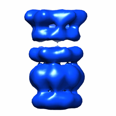 400_5923.gif
 80_5923.gif
 http://ftp.pdbj.org/pub/emdb/structures/EMD-5923


 Sample components
 Processing
 Electron microscopy