 Movie
Movie Controller

[English] 日本語
 Yorodumi
- EMDB-5828: Architecture of a dsDNA viral capsid in complex with its maturati... -
+ Open data

- Basic information
| Entry | Database: EMDB / ID: EMD-5828 | |||||||||
|---|---|---|---|---|---|---|---|---|---|---|
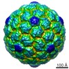
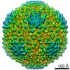
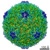
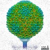
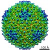
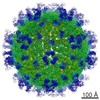
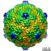
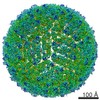
| Title | Architecture of a dsDNA viral capsid in complex with its maturation protease | |||||||||
 Map data Map data | Prohead-1 without protease (before sharpening). A negative temperature factor of -300 A2 or -700 A2 should be applied to better visualize the features of the scaffolding domains or the coat subunit shell, respectively. | |||||||||
 Sample Sample |
| |||||||||
 Keywords Keywords | Bacteriophage HK97 / virus maturation / electron cryo-microscopy / scaffolding proteins | |||||||||
| Biological species |  Enterobacteria phage HK97 (virus) Enterobacteria phage HK97 (virus) | |||||||||
| Method | single particle reconstruction / cryo EM / Resolution: 7.8 Å | |||||||||
 Authors Authors | Veesler D / Khayat R / Krishnamurthy S / Snijder J / Huang RK / Heck AJR / Anand GS / Johnson JE | |||||||||
 Citation Citation | Journal: Structure / Year: 2014 Title: Architecture of a dsDNA viral capsid in complex with its maturation protease. Authors: David Veesler / Reza Khayat / Srinath Krishnamurthy / Joost Snijder / Rick K Huang / Albert J R Heck / Ganesh S Anand / John E Johnson /    Abstract: Most double-stranded DNA (dsDNA) viruses, including bacteriophages and herpesviruses, rely on a staged assembly process of capsid formation. A viral protease is required for many of them to ...Most double-stranded DNA (dsDNA) viruses, including bacteriophages and herpesviruses, rely on a staged assembly process of capsid formation. A viral protease is required for many of them to disconnect scaffolding domains/proteins from the capsid shell, therefore priming the maturation process. We used the bacteriophage HK97 as a model system to decipher the molecular mechanisms underlying the recruitment of the maturation protease by the assembling procapsid and the influence exerted onto the latter. Comparisons of the procapsid with and without protease using single-particle cryoelectron microscopy reconstructions, hydrogen/deuterium exchange coupled to mass spectrometry, and native mass spectrometry demonstrated that the protease interacts with the scaffolding domains within the procapsid interior and stabilizes them as well as the whole particle. The results suggest that the thermodynamic consequences of protease packaging are to shift the equilibrium between isolated coat subunit capsomers and procapsid in favor of the latter by stabilizing the assembled particle before making the process irreversible through proteolysis of the scaffolding domains. | |||||||||
| History |
|
- Structure visualization
| Movie |
 Movie viewer Movie viewer |
|---|---|
| Structure viewer | EM map: SurfViewMolmilJmol/JSmol |
| Supplemental images |

- Downloads & links
-EMDB archive
| Map data | emd_5828.map.gz | 17.9 MB | EMDB map data format | |
|---|---|---|---|---|
| Header (meta data) | emd-5828-v30.xmlemd-5828.xml | 9.7 KB 9.7 KB | Display Display | EMDB header |
| Images |  400_5828.gif 400_5828.gif 80_5828.gif 80_5828.gif | 82.9 KB 5.2 KB | ||
| Archive directory |  http://ftp.pdbj.org/pub/emdb/structures/EMD-5828ftp://ftp.pdbj.org/pub/emdb/structures/EMD-5828 http://ftp.pdbj.org/pub/emdb/structures/EMD-5828ftp://ftp.pdbj.org/pub/emdb/structures/EMD-5828 | HTTPS FTP |
-Validation report
| Summary document | emd_5828_validation.pdf.gz | 78.9 KB | Display | EMDB validaton report |
|---|---|---|---|---|
| Full document | emd_5828_full_validation.pdf.gz | 78 KB | Display | |
| Data in XML | emd_5828_validation.xml.gz | 494 B | Display | |
| Arichive directory | https://ftp.pdbj.org/pub/emdb/validation_reports/EMD-5828ftp://ftp.pdbj.org/pub/emdb/validation_reports/EMD-5828 | HTTPS FTP |
-Related structure data

-Links
| EMDB pages | EMDB (EBI/PDBe) / EMDataResource |
|---|
-Map
| File | Download / File: emd_5828.map.gz / Format: CCP4 / Size: 300.3 MB / Type: IMAGE STORED AS FLOATING POINT NUMBER (4 BYTES) | ||||||||||||||||||||||||||||||||||||||||||||||||||||||||||||||||||||
|---|---|---|---|---|---|---|---|---|---|---|---|---|---|---|---|---|---|---|---|---|---|---|---|---|---|---|---|---|---|---|---|---|---|---|---|---|---|---|---|---|---|---|---|---|---|---|---|---|---|---|---|---|---|---|---|---|---|---|---|---|---|---|---|---|---|---|---|---|---|
| Annotation | Prohead-1 without protease (before sharpening). A negative temperature factor of -300 A2 or -700 A2 should be applied to better visualize the features of the scaffolding domains or the coat subunit shell, respectively. | ||||||||||||||||||||||||||||||||||||||||||||||||||||||||||||||||||||
| Voxel size | X=Y=Z: 2.75 Å | ||||||||||||||||||||||||||||||||||||||||||||||||||||||||||||||||||||
| Density |
| ||||||||||||||||||||||||||||||||||||||||||||||||||||||||||||||||||||
| Symmetry | Space group: 1 | ||||||||||||||||||||||||||||||||||||||||||||||||||||||||||||||||||||
| Details | EMDB XML:
CCP4 map header:
| ||||||||||||||||||||||||||||||||||||||||||||||||||||||||||||||||||||
-Supplemental data
- Sample components
-Entire : HK97 Prohead-1 without protease
| Entire | Name: HK97 Prohead-1 without protease |
|---|---|
| Components |
|
-Supramolecule #1000: HK97 Prohead-1 without protease
| Supramolecule | Name: HK97 Prohead-1 without protease / type: sample / ID: 1000 / Oligomeric state: icosahedral / Number unique components: 1 |
|---|---|
| Molecular weight | Experimental: 17.9 MDa / Theoretical: 17.7 MDa / Method: Native mass spectrometry |
-Supramolecule #1: Enterobacteria phage HK97
| Supramolecule | Name: Enterobacteria phage HK97 / type: virus / ID: 1 / NCBI-ID: 37554 / Sci species name: Enterobacteria phage HK97 / Sci species strain: HK97 / Database: NCBI / Virus type: VIRUS-LIKE PARTICLE / Virus isolate: STRAIN / Virus enveloped: No / Virus empty: Yes |
|---|---|
| Host (natural) | Organism:  |
| Virus shell | Shell ID: 1 / Name: gp5 / Diameter: 540 Å / T number (triangulation number): 7 |
-Experimental details
-Structure determination
| Method | cryo EM |
|---|---|
 Processing Processing | single particle reconstruction |
| Aggregation state | particle |
-Sample preparation
| Buffer | pH: 7.5 / Details: 10 mM Tris, 40 mM monosodium glutamate |
|---|---|
| Grid | Details: C-flat |
| Vitrification | Cryogen name: ETHANE / Chamber temperature: 94 K / Instrument: FEI VITROBOT MARK II |
- Electron microscopy
| Microscope | FEI TECNAI F20 |
|---|---|
| Temperature | Average: 90 K |
| Date | Nov 18, 2008 |
| Image recording | Category: CCD / Film or detector model: GATAN ULTRASCAN 4000 (4k x 4k) / Number real images: 208 / Average electron dose: 16 e/Å2 |
| Electron beam | Acceleration voltage: 120 kV / Electron source:  FIELD EMISSION GUN FIELD EMISSION GUN |
| Electron optics | Calibrated magnification: 109489 / Illumination mode: FLOOD BEAM / Imaging mode: BRIGHT FIELD / Cs: 2.0 mm / Nominal defocus max: 2.749 µm / Nominal defocus min: 0.8 µm |
| Sample stage | Specimen holder: Liquid Nitrogen cooled / Specimen holder model: GATAN LIQUID NITROGEN |
| Experimental equipment |  Model: Tecnai F20 / Image courtesy: FEI Company |
-Image processing
| CTF correction | Details: Each particle |
|---|---|
| Final reconstruction | Algorithm: OTHER / Resolution.type: BY AUTHOR / Resolution: 7.8 Å / Resolution method: OTHER / Software - Name: Frealign / Details: Resolution-limited refinement to 10 A / Number images used: 5256 |
