 Movie
Movie Controller


+ Open data

- Basic information
| Entry | Database: EMDB / ID: EMD-5786 | |||||||||
|---|---|---|---|---|---|---|---|---|---|---|
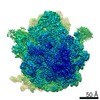
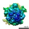
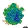
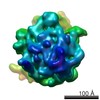
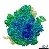
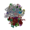
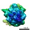
| Title | EttA-bound E. coli 70S ribosome complex | |||||||||
 Map data Map data | Reconstruction of E. coli 70S ribosome complex containing EttA-EQ2 mutant protein | |||||||||
 Sample Sample |
| |||||||||
 Keywords Keywords | protein translation regulation / ABC-F protein family / ribosome / cryo-EM / single-molecule FRET / YjjK | |||||||||
| Function / homology |  Function and homology information Function and homology informationnegative regulation of translational elongation / Hydrolases; Acting on acid anhydrides; In phosphorus-containing anhydrides / ribosome binding / tRNA binding / rRNA binding / translation / ATP hydrolysis activity / ATP binding / cytoplasm Similarity search - Function | |||||||||
| Biological species |  | |||||||||
| Method | single particle reconstruction / cryo EM / Resolution: 7.7 Å | |||||||||
 Authors Authors | Chen B / Boel G / Hashem Y / Ning W / Fei J / Wang C / Gonzalez RL / Hunt JF / Frank J | |||||||||
 Citation Citation | Journal: Nat Struct Mol Biol / Year: 2014 Title: The ABC-F protein EttA gates ribosome entry into the translation elongation cycle. Authors: Grégory Boël / Paul C Smith / Wei Ning / Michael T Englander / Bo Chen / Yaser Hashem / Anthony J Testa / Jeffrey J Fischer / Hans-Joachim Wieden / Joachim Frank / Ruben L Gonzalez / John F Hunt /   Abstract: ABC-F proteins have evaded functional characterization even though they compose one of the most widely distributed branches of the ATP-binding cassette (ABC) superfamily. Herein, we demonstrate that ...ABC-F proteins have evaded functional characterization even though they compose one of the most widely distributed branches of the ATP-binding cassette (ABC) superfamily. Herein, we demonstrate that YjjK, the most prevalent eubacterial ABC-F protein, gates ribosome entry into the translation elongation cycle through a nucleotide-dependent interaction sensitive to ATP/ADP ratio. Accordingly, we rename this protein energy-dependent translational throttle A (EttA). We determined the crystal structure of Escherichia coli EttA and used it to design mutants for biochemical studies including enzymological assays of the initial steps of protein synthesis. These studies suggest that EttA may regulate protein synthesis in energy-depleted cells, which have a low ATP/ADP ratio. Consistently with this inference, EttA-deleted cells exhibit a severe fitness defect in long-term stationary phase. These studies demonstrate that an ABC-F protein regulates protein synthesis via a new mechanism sensitive to cellular energy status. | |||||||||
| History |
|
- Structure visualization
| Movie |
Movie viewer |
|---|---|
| Structure viewer | EM map: SurfViewMolmilJmol/JSmol |
| Supplemental images |

- Downloads & links
-EMDB archive
| Map data | emd_5786.map.gz | 2.2 MB | EMDB map data format | |
|---|---|---|---|---|
| Header (meta data) | emd-5786-v30.xmlemd-5786.xml | 15.2 KB 15.2 KB | Display Display | EMDB header |
| Images | 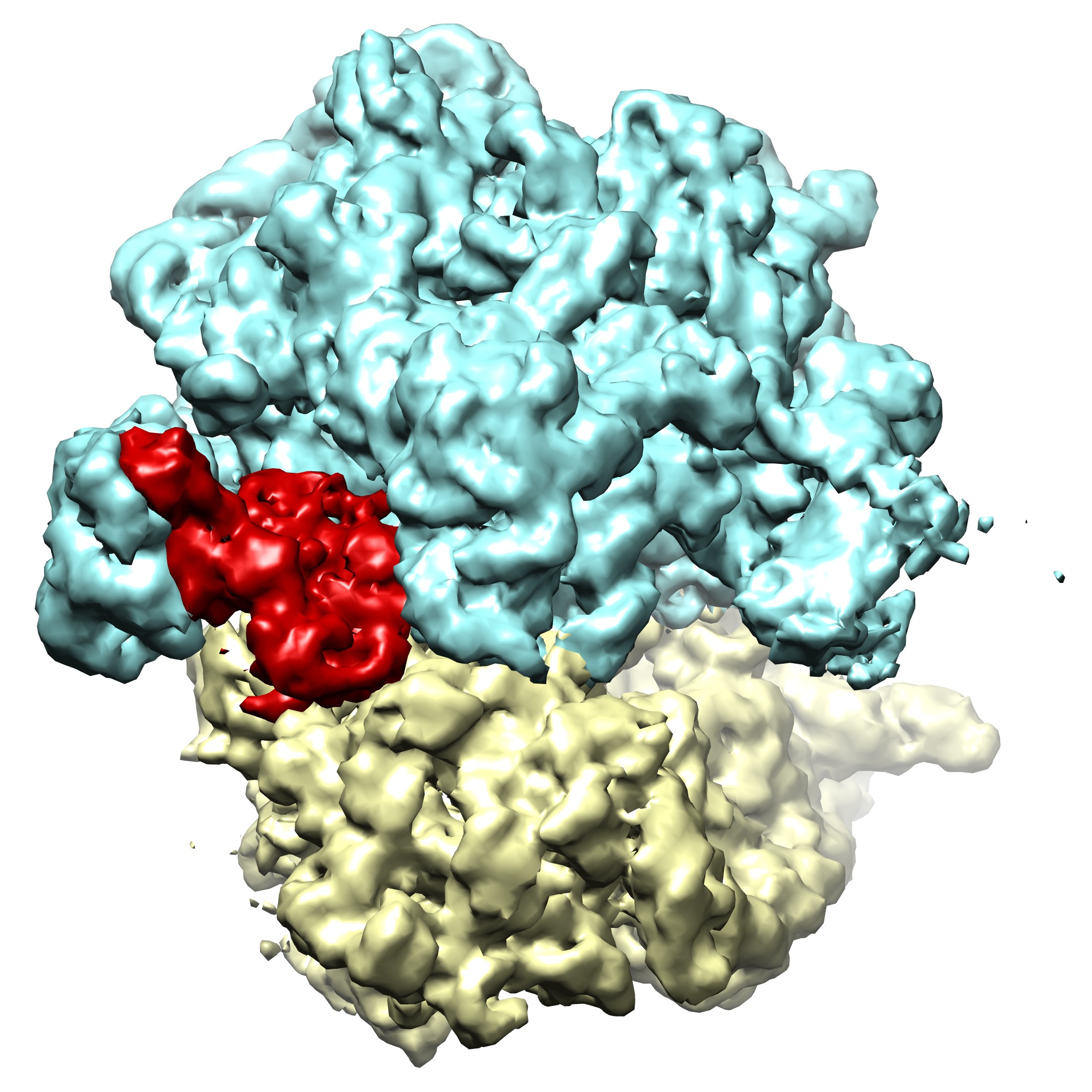 emd_5786.jpg emd_5786.jpg | 512.7 KB | ||
| Archive directory |  http://ftp.pdbj.org/pub/emdb/structures/EMD-5786ftp://ftp.pdbj.org/pub/emdb/structures/EMD-5786 http://ftp.pdbj.org/pub/emdb/structures/EMD-5786ftp://ftp.pdbj.org/pub/emdb/structures/EMD-5786 | HTTPS FTP |
-Validation report
| Summary document | emd_5786_validation.pdf.gz | 78.6 KB | Display | EMDB validaton report |
|---|---|---|---|---|
| Full document | emd_5786_full_validation.pdf.gz | 77.7 KB | Display | |
| Data in XML | emd_5786_validation.xml.gz | 494 B | Display | |
| Arichive directory | https://ftp.pdbj.org/pub/emdb/validation_reports/EMD-5786ftp://ftp.pdbj.org/pub/emdb/validation_reports/EMD-5786 | HTTPS FTP |
-Related structure data
| Related structure data |  5784C  5785C  5841C 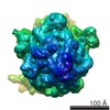 5842C 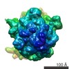 5843C  3j5sC C: citing same article ( |
|---|---|
| Similar structure data |
-Links
| EMDB pages | EMDB (EBI/PDBe) / EMDataResource |
|---|---|
| Related items in Molecule of the Month |


-Map
| File | Download / File: emd_5786.map.gz / Format: CCP4 / Size: 9 MB / Type: IMAGE STORED AS FLOATING POINT NUMBER (4 BYTES) | ||||||||||||||||||||||||||||||||||||||||||||||||||||||||||||||||||||
|---|---|---|---|---|---|---|---|---|---|---|---|---|---|---|---|---|---|---|---|---|---|---|---|---|---|---|---|---|---|---|---|---|---|---|---|---|---|---|---|---|---|---|---|---|---|---|---|---|---|---|---|---|---|---|---|---|---|---|---|---|---|---|---|---|---|---|---|---|---|
| Annotation | Reconstruction of E. coli 70S ribosome complex containing EttA-EQ2 mutant protein | ||||||||||||||||||||||||||||||||||||||||||||||||||||||||||||||||||||
| Voxel size | X=Y=Z: 2.7116 Å | ||||||||||||||||||||||||||||||||||||||||||||||||||||||||||||||||||||
| Density |
| ||||||||||||||||||||||||||||||||||||||||||||||||||||||||||||||||||||
| Symmetry | Space group: 1 | ||||||||||||||||||||||||||||||||||||||||||||||||||||||||||||||||||||
| Details | EMDB XML:
CCP4 map header:
| ||||||||||||||||||||||||||||||||||||||||||||||||||||||||||||||||||||
-Supplemental data
- Sample components
-Entire : E. coli 70S ribosome complex 70S-EttA_EQ2
| Entire | Name: E. coli 70S ribosome complex 70S-EttA_EQ2 |
|---|---|
| Components |
|
-Supramolecule #1000: E. coli 70S ribosome complex 70S-EttA_EQ2
| Supramolecule | Name: E. coli 70S ribosome complex 70S-EttA_EQ2 / type: sample / ID: 1000 / Number unique components: 2 |
|---|
-Supramolecule #1: 70S ribosome
| Supramolecule | Name: 70S ribosome / type: complex / ID: 1 / Recombinant expression: No / Database: NCBI / Ribosome-details: ribosome-prokaryote: ALL |
|---|---|
| Ref GO | 0: GO:0042255 |
| Source (natural) | Organism: |
| Molecular weight | Experimental: 2.7 MDa |
-Macromolecule #1: Energy-dependent Translational Throttle A (EttA)
| Macromolecule | Name: Energy-dependent Translational Throttle A (EttA) / type: protein_or_peptide / ID: 1 / Name.synonym: YjjK / Recombinant expression: Yes |
|---|---|
| Source (natural) | Organism: |
| Molecular weight | Theoretical: 60 KDa |
| Recombinant expression | Organism: |
| Sequence | UniProtKB: Energy-dependent translational throttle protein EttA |
-Experimental details
-Structure determination
| Method | cryo EM |
|---|---|
 Processing Processing | single particle reconstruction |
| Aggregation state | particle |
-Sample preparation
| Concentration | 1.2 mg/mL |
|---|---|
| Buffer | pH: 6.9 Details: 50 mM Tris acetate, 100 mM KCl, 5 mM NH4OAc, 3.5 mM Mg(OAc)2, 0.5 mM Ca(OAc)2, 0.1 mM EDTA, 1 mM spermidine, 5 mM putrescine, 6 mM 2-mercaptoethanol, 0.5 mM Mg-ATP |
| Grid | Details: Quantifoil R2/4 300 mesh Cu EM grid, coated with thin carbon film, glow discharged in H2/O2 |
| Vitrification | Cryogen name: ETHANE / Chamber humidity: 100 % / Chamber temperature: 80 K / Instrument: FEI VITROBOT MARK IV Method: Wait time 30 sec, blot time 8 sec, at 4 degrees Celsius |
- Electron microscopy #1
| Microscopy ID | 1 |
|---|---|
| Microscope | FEI TECNAI F20 |
| Temperature | Average: 80 K |
| Details | Low dose |
| Date | Apr 5, 2011 |
| Image recording | Category: CCD / Film or detector model: GATAN ULTRASCAN 4000 (4k x 4k) / Number real images: 574 / Average electron dose: 17 e/Å2 Details: Used the automatic image collection program Leginon |
| Electron beam | Acceleration voltage: 200 kV / Electron source:  FIELD EMISSION GUN FIELD EMISSION GUN |
| Electron optics | Calibrated magnification: 110637 / Illumination mode: FLOOD BEAM / Imaging mode: BRIGHT FIELD / Cs: 2.0 mm / Nominal defocus max: 3.5 µm / Nominal defocus min: 1.2 µm / Nominal magnification: 80000 |
| Sample stage | Specimen holder: Single tilt cryoholder, liquid Nitrogen cooled Specimen holder model: GATAN LIQUID NITROGEN |
| Experimental equipment |  Model: Tecnai F20 / Image courtesy: FEI Company |
-Electron microscopy #2
| Microscopy ID | 2 |
|---|---|
| Microscope | FEI TECNAI F20 |
| Temperature | Average: 80 K |
| Details | Low dose |
| Date | Jun 6, 2011 |
| Image recording | Category: CCD / Film or detector model: GATAN ULTRASCAN 4000 (4k x 4k) / Number real images: 1816 / Average electron dose: 17 e/Å2 Details: Used the automatic image collection program Leginon |
| Electron beam | Acceleration voltage: 200 kV / Electron source: FIELD EMISSION GUN |
| Electron optics | Calibrated magnification: 110637 / Illumination mode: FLOOD BEAM / Imaging mode: BRIGHT FIELD / Cs: 2.0 mm / Nominal defocus max: 3.5 µm / Nominal defocus min: 1.2 µm / Nominal magnification: 80000 |
| Sample stage | Specimen holder: Single tilt cryoholder, liquid Nitrogen cooled Specimen holder model: GATAN LIQUID NITROGEN |
| Experimental equipment | Model: Tecnai F20 / Image courtesy: FEI Company |
-Image processing
| Details | The particles were selected via automatic particle picking followed by visual verification. 3D classification and refinement were performed using RELION. |
|---|---|
| CTF correction | Details: Each micrograph |
| Final reconstruction | Resolution.type: BY AUTHOR / Resolution: 7.7 Å / Resolution method: OTHER / Software - Name: SPIDER, RELION / Details: Subset after RELION 3D classification / Number images used: 33889 |
