 Movie
Movie Controller

[English] 日本語
 Yorodumi
- EMDB-5762: Electron cryo-microscopy of four-stranded TubZ from B. thuringiensis -
+ Open data

- Basic information
| Entry | Database: EMDB / ID: EMD-5762 | |||||||||
|---|---|---|---|---|---|---|---|---|---|---|
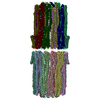
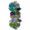
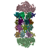
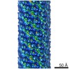
| Title | Electron cryo-microscopy of four-stranded TubZ from B. thuringiensis | |||||||||
 Map data Map data | Reconstruction of four-stranded TubZ filament grown in the presence of GTP | |||||||||
 Sample Sample |
| |||||||||
 Keywords Keywords | plasmid segregation /  tubulin family / bacterial cytoskeleton tubulin family / bacterial cytoskeleton | |||||||||
| Function / homology |  Function and homology information Function and homology informationplasmid partitioning / Hydrolases; Acting on acid anhydrides; Acting on GTP to facilitate cellular and subcellular movement / GTPase activity / GTP binding / identical protein binding / metal ion binding / cytoplasmSimilarity search - Function | |||||||||
| Biological species |  Bacillus thuringiensis (bacteria) Bacillus thuringiensis (bacteria) | |||||||||
| Method | helical reconstruction / cryo EM / Resolution: 6.9 Å | |||||||||
 Authors Authors | Montabana EA / Agard DA | |||||||||
 Citation Citation | Journal: Proc Natl Acad Sci U S A / Year: 2014 Title: Bacterial tubulin TubZ-Bt transitions between a two-stranded intermediate and a four-stranded filament upon GTP hydrolysis. Authors: Elizabeth A Montabana / David A Agard /  Abstract: Cytoskeletal filaments form diverse superstructures that are highly adapted for specific functions. The recently discovered TubZ subfamily of tubulins is involved in type III plasmid partitioning ...Cytoskeletal filaments form diverse superstructures that are highly adapted for specific functions. The recently discovered TubZ subfamily of tubulins is involved in type III plasmid partitioning systems, facilitating faithful segregation of low copy-number plasmids during bacterial cell division. One such protein, TubZ-Bt, is found on the large pBtoxis plasmid in Bacillus thuringiensis, and interacts via its extended C terminus with a DNA adaptor protein TubR. Here, we use cryo-electron microscopy to determine the structure of TubZ-Bt filaments and light scattering to explore their mechanism of polymerization. Surprisingly, we find that the helical filament architecture is remarkably sensitive to nucleotide state, changing from two-stranded to four-stranded depending on the ability of TubZ-Bt to hydrolyze GTP. We present pseudoatomic models of both the two- and four-protofilament forms based on cryo-electron microscopy reconstructions (10.8 Å and 6.9 Å, respectively) of filaments formed under different nucleotide states. These data lead to a model in which the two-stranded filament is a necessary intermediate along the pathway to formation of the four-stranded filament. Such nucleotide-directed structural polymorphism is to our knowledge an unprecedented mechanism for the formation of polar filaments. | |||||||||
| History |
|
- Structure visualization
| Movie |
Movie viewer |
|---|---|
| Structure viewer | EM map: SurfViewMolmilJmol/JSmol |
| Supplemental images |

- Downloads & links
-EMDB archive
| Map data | emd_5762.map.gz | 20.9 MB | EMDB map data format | |
|---|---|---|---|---|
| Header (meta data) | emd-5762-v30.xmlemd-5762.xml | 10.5 KB 10.5 KB | Display Display | EMDB header |
| Images | 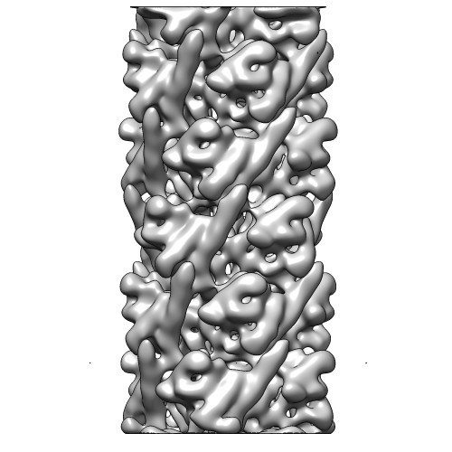 emd_5762.jpg emd_5762.jpg | 46.9 KB | ||
| Archive directory |  http://ftp.pdbj.org/pub/emdb/structures/EMD-5762ftp://ftp.pdbj.org/pub/emdb/structures/EMD-5762 http://ftp.pdbj.org/pub/emdb/structures/EMD-5762ftp://ftp.pdbj.org/pub/emdb/structures/EMD-5762 | HTTPS FTP |
-Related structure data
| Related structure data |  3j4sMC  5763C  3j4tC M: atomic model generated by this map C: citing same article ( |
|---|---|
| Similar structure data |


-Links
| EMDB pages | EMDB (EBI/PDBe) / EMDataResource |
|---|---|
| Related items in Molecule of the Month |


-Map
| File | Download / File: emd_5762.map.gz / Format: CCP4 / Size: 62.5 MB / Type: IMAGE STORED AS FLOATING POINT NUMBER (4 BYTES) | ||||||||||||||||||||||||||||||||||||||||||||||||||||||||||||||||||||
|---|---|---|---|---|---|---|---|---|---|---|---|---|---|---|---|---|---|---|---|---|---|---|---|---|---|---|---|---|---|---|---|---|---|---|---|---|---|---|---|---|---|---|---|---|---|---|---|---|---|---|---|---|---|---|---|---|---|---|---|---|---|---|---|---|---|---|---|---|---|
| Annotation | Reconstruction of four-stranded TubZ filament grown in the presence of GTP | ||||||||||||||||||||||||||||||||||||||||||||||||||||||||||||||||||||
| Voxel size | X=Y=Z: 0.94 Å | ||||||||||||||||||||||||||||||||||||||||||||||||||||||||||||||||||||
| Density |
| ||||||||||||||||||||||||||||||||||||||||||||||||||||||||||||||||||||
| Symmetry | Space group: 1 | ||||||||||||||||||||||||||||||||||||||||||||||||||||||||||||||||||||
| Details | EMDB XML:
CCP4 map header:
| ||||||||||||||||||||||||||||||||||||||||||||||||||||||||||||||||||||
-Supplemental data
- Sample components
-Entire : Untagged full length TubZ from pBtoxis in Bacillus thuringiensis
| Entire | Name: Untagged full length TubZ from pBtoxis in Bacillus thuringiensis |
|---|---|
| Components |
|
-Supramolecule #1000: Untagged full length TubZ from pBtoxis in Bacillus thuringiensis
| Supramolecule | Name: Untagged full length TubZ from pBtoxis in Bacillus thuringiensis type: sample / ID: 1000 Oligomeric state: Helical filament with four protofilament strands Number unique components: 1 |
|---|
-Macromolecule #1: ftsZ/tubulin-related protein
| Macromolecule | Name: ftsZ/tubulin-related protein / type: protein_or_peptide / ID: 1 / Name.synonym: TubZ-Bt, TubZ / Oligomeric state: helical four-stranded filament / Recombinant expression: Yes |
|---|---|
| Source (natural) | Organism: Bacillus thuringiensis (bacteria) / Strain: serovar israelensis |
| Molecular weight | Experimental: 54.3729 KDa |
| Recombinant expression | Organism: Escherichia coli (E. coli) |
| Sequence | UniProtKB: Tubulin-like protein TubZ / InterPro: Tubulin/FtsZ, GTPase domain |
-Experimental details
-Structure determination
| Method | cryo EM |
|---|---|
 Processing Processing | helical reconstruction |
| Aggregation state | filament |
-Sample preparation
| Buffer | pH: 7.7 Details: 100 mM potassium acetate, 5 mM magnesium acetate, 50 mM HEPES |
|---|---|
| Grid | Details: 400 mesh copper grid with holey carbon support, glow discharged |
| Vitrification | Cryogen name: ETHANE / Chamber humidity: 100 % / Instrument: FEI VITROBOT MARK III Timed resolved state: GTP added to TubZ protein and incubated ~6 minutes before placing on grid. Method: Blot 4.5 seconds before plunging. |
- Electron microscopy
| Microscope | FEI TECNAI F20 |
|---|---|
| Electron beam | Acceleration voltage: 200 kV / Electron source: FIELD EMISSION GUN |
| Electron optics | Illumination mode: FLOOD BEAM / Imaging mode: BRIGHT FIELDBright-field microscopy / Cs: 2.2 mm / Nominal defocus max: 3.5 µm / Nominal defocus min: 0.8 µm / Nominal magnification: 80000 |
| Sample stage | Specimen holder model: OTHER |
| Alignment procedure | Legacy - Astigmatism: Astigmatism corrected at 250,000 times magnification |
| Date | Apr 22, 2011 |
| Image recording | Category: CCD / Film or detector model: TVIPS TEMCAM-F816 (8k x 8k) / Number real images: 414 / Average electron dose: 20 e/Å2 |
| Experimental equipment |  Model: Tecnai F20 / Image courtesy: FEI Company |
-Image processing
| CTF correction | Details: Whole micrograph |
|---|---|
| Final reconstruction | Applied symmetry - Helical parameters - Δz: 43.54512 Å Applied symmetry - Helical parameters - Δ&Phi: 31.78843 ° Algorithm: OTHER / Resolution.type: BY AUTHOR / Resolution: 6.9 Å / Resolution method: FSC 0.143 CUT-OFF / Software - Name: Spider Details: Final Map has been low-pass filtered to 7 Angstrom and high-pass filtered to 30 Angstrom. A B-factor of -309 Angstrom was applied using the program bfactor. A cylindrical mask of radius ~80 ...Details: Final Map has been low-pass filtered to 7 Angstrom and high-pass filtered to 30 Angstrom. A B-factor of -309 Angstrom was applied using the program bfactor. A cylindrical mask of radius ~80 Angstrom has been applied. |
| Details | The particles were aligned using IHRSR. |

