 Movie
Movie Controller


+ Open data

- Basic information
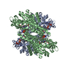
| Entry | Database: EMDB / ID: EMD-5739 | |||||||||
|---|---|---|---|---|---|---|---|---|---|---|
| Title | Electron microscopy map of the T6SS TssK component | |||||||||
 Map data Map data | EM Reconstruction of TssK | |||||||||
 Sample Sample |
| |||||||||
 Keywords Keywords | TssK / Type VI secretion / Hcp / sheath assembly / EM / single-particle / three-fold | |||||||||
| Function / homology | cellular_component / biological_process / molecular_function / Type VI secretion system TssK / Type VI secretion system TssK / Bacterial Type VI secretion, VC_A0110, EvfL, ImpJ, VasE / Type VI secretion system protein TssK Function and homology information Function and homology information | |||||||||
| Biological species |  | |||||||||
| Method | single particle reconstruction / negative staining / Resolution: 26.0 Å | |||||||||
 Authors Authors | Zoued A / Durand E / Bebeacua C / Brunet YR / Douzi B / Cambillau C / Cascales E / Journet L | |||||||||
 Citation Citation | Journal: J Biol Chem / Year: 2013 Title: TssK is a trimeric cytoplasmic protein interacting with components of both phage-like and membrane anchoring complexes of the type VI secretion system. Authors: Abdelrahim Zoued / Eric Durand / Cecilia Bebeacua / Yannick R Brunet / Badreddine Douzi / Christian Cambillau / Eric Cascales / Laure Journet /  Abstract: The Type VI secretion system (T6SS) is a macromolecular machine that mediates bacteria-host or bacteria-bacteria interactions. The T6SS core apparatus assembles from 13 proteins that form two sub- ...The Type VI secretion system (T6SS) is a macromolecular machine that mediates bacteria-host or bacteria-bacteria interactions. The T6SS core apparatus assembles from 13 proteins that form two sub-assemblies: a phage-like complex and a trans-envelope complex. The Hcp, VgrG, TssE, and TssB/C subunits are structurally and functionally related to components of the tail of contractile bacteriophages. This phage-like structure is thought to be anchored to the membrane by a trans-envelope complex composed of the TssJ, TssL, and TssM proteins. However, how the two sub-complexes are connected remains unknown. Here we identify TssK, a protein that establishes contacts with the two T6SS sub-complexes through direct interactions with TssL, Hcp, and TssC. TssK is a cytoplasmic protein assembling trimers that display a three-armed shape, as revealed by TEM and SAXS analyses. Fluorescence microscopy experiments further demonstrate the requirement of TssK for sheath assembly. Our results suggest a central role for TssK by linking both complexes during T6SS assembly. | |||||||||
| History |
|
- Structure visualization
| Movie |
Movie viewer |
|---|---|
| Structure viewer | EM map: SurfViewMolmilJmol/JSmol |
| Supplemental images |
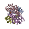
 UCSF Chimera
- Downloads & links
-EMDB archive
| Map data | emd_5739.map.gz | 944 KB | EMDB map data format | |
|---|---|---|---|---|
| Header (meta data) | emd-5739-v30.xmlemd-5739.xml | 10.4 KB 10.4 KB | Display Display | EMDB header |
| Images | 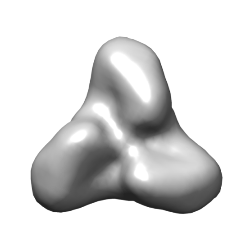 emd_5739.png emd_5739.png | 42.8 KB | ||
| Archive directory |  http://ftp.pdbj.org/pub/emdb/structures/EMD-5739ftp://ftp.pdbj.org/pub/emdb/structures/EMD-5739 http://ftp.pdbj.org/pub/emdb/structures/EMD-5739ftp://ftp.pdbj.org/pub/emdb/structures/EMD-5739 | HTTPS FTP |
-Validation report
| Summary document | emd_5739_validation.pdf.gz | 78.7 KB | Display | EMDB validaton report |
|---|---|---|---|---|
| Full document | emd_5739_full_validation.pdf.gz | 77.8 KB | Display | |
| Data in XML | emd_5739_validation.xml.gz | 494 B | Display | |
| Arichive directory | https://ftp.pdbj.org/pub/emdb/validation_reports/EMD-5739ftp://ftp.pdbj.org/pub/emdb/validation_reports/EMD-5739 | HTTPS FTP |
-Related structure data
| Similar structure data |
|---|


-Links
| EMDB pages | EMDB (EBI/PDBe) / EMDataResource |
|---|
-Map
| File | Download / File: emd_5739.map.gz / Format: CCP4 / Size: 1.4 MB / Type: IMAGE STORED AS FLOATING POINT NUMBER (4 BYTES) | ||||||||||||||||||||||||||||||||||||||||||||||||||||||||||||||||||||
|---|---|---|---|---|---|---|---|---|---|---|---|---|---|---|---|---|---|---|---|---|---|---|---|---|---|---|---|---|---|---|---|---|---|---|---|---|---|---|---|---|---|---|---|---|---|---|---|---|---|---|---|---|---|---|---|---|---|---|---|---|---|---|---|---|---|---|---|---|---|
| Annotation | EM Reconstruction of TssK | ||||||||||||||||||||||||||||||||||||||||||||||||||||||||||||||||||||
| Voxel size | X=Y=Z: 3.5 Å | ||||||||||||||||||||||||||||||||||||||||||||||||||||||||||||||||||||
| Density |
| ||||||||||||||||||||||||||||||||||||||||||||||||||||||||||||||||||||
| Symmetry | Space group: 1 | ||||||||||||||||||||||||||||||||||||||||||||||||||||||||||||||||||||
| Details | EMDB XML:
CCP4 map header:
| ||||||||||||||||||||||||||||||||||||||||||||||||||||||||||||||||||||
-Supplemental data
- Sample components
-Entire : Purified TssK sample
| Entire | Name: Purified TssK sample |
|---|---|
| Components |
|
-Supramolecule #1000: Purified TssK sample
| Supramolecule | Name: Purified TssK sample / type: sample / ID: 1000 / Details: The sample was monodisperse. / Oligomeric state: 3 / Number unique components: 1 |
|---|---|
| Molecular weight | Experimental: 160 KDa / Method: Analytical size exclusion chromatography analysis |
-Macromolecule #1: Type VI secretion system protein TssK
| Macromolecule | Name: Type VI secretion system protein TssK / type: protein_or_peptide / ID: 1 / Name.synonym: TssK / Number of copies: 1 / Oligomeric state: Trimer / Recombinant expression: Yes |
|---|---|
| Source (natural) | Organism: |
| Molecular weight | Experimental: 160 KDa |
| Recombinant expression | Organism: |
| Sequence | UniProtKB: Type VI secretion system protein TssK GO: biological_process, molecular_function, cellular_component InterPro: Type VI secretion system TssK |
-Experimental details
-Structure determination
| Method | negative staining |
|---|---|
 Processing Processing | single particle reconstruction |
| Aggregation state | particle |
-Sample preparation
| Concentration | 0.05 mg/mL |
|---|---|
| Buffer | pH: 8 Details: HBS-EP buffer (10mM HEPES, 150mM NaCl, 3mM EDTA, 0.005% Polysorbate 20) |
| Staining | Type: NEGATIVE Details: Purified protein was immobilized on a glow- discharged carbon grid by incubation for 1 minute, and then stained with 2% uranyl acetate for 10 seconds. |
| Grid | Details: 300 mesh copper grid with thin carbon support, glow discharged for 20 seconds |
| Vitrification | Cryogen name: NONE / Instrument: OTHER |
- Electron microscopy
| Microscope | FEI TECNAI SPIRIT |
|---|---|
| Date | Nov 1, 2012 |
| Image recording | Category: CCD / Film or detector model: GENERIC CCD / Number real images: 10 / Average electron dose: 10 e/Å2 / Details: CCD images |
| Electron beam | Acceleration voltage: 120 kV / Electron source: LAB6 |
| Electron optics | Illumination mode: FLOOD BEAM / Imaging mode: BRIGHT FIELD / Cs: 2 mm / Nominal defocus max: 3.0 µm / Nominal defocus min: 2.0 µm / Nominal magnification: 60000 |
| Sample stage | Specimen holder: Room temperature holder / Specimen holder model: SIDE ENTRY, EUCENTRIC |
| Experimental equipment |  Model: Tecnai Spirit / Image courtesy: FEI Company |
-Image processing
| Details | The particles were submitted to maximum likelihood classification and alignment. |
|---|---|
| Final reconstruction | Algorithm: OTHER / Resolution.type: BY AUTHOR / Resolution: 26.0 Å / Resolution method: OTHER / Software - Name: EMAN, SPIDER, XMIPP / Number images used: 5000 |
| Final angle assignment | Details: ML |
| Final two d classification | Number classes: 500 |
