 Movie
Movie Controller

[English] 日本語
 Yorodumi
- EMDB-5626: Molecular Architecture of the ATP-Dependent Chromatin Remodeling ... -
+ Open data

- Basic information
| Entry | Database: EMDB / ID: EMD-5626 | |||||||||
|---|---|---|---|---|---|---|---|---|---|---|
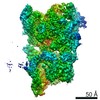
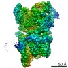
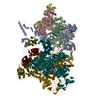
| Title | Molecular Architecture of the ATP-Dependent Chromatin Remodeling Complex SWR1 by 3 Dimensional Electron Microscopy | |||||||||
 Map data Map data | 3D Cryo-negative EM structure of SWR1 | |||||||||
 Sample Sample |
| |||||||||
 Keywords Keywords | SWR1 / ATP-dependent chromatin remodeling /  nucleosome / H2A.Z / Rvb1 / Rvb2 / AAA+ ATPases / 3 dimensional electron microscopy nucleosome / H2A.Z / Rvb1 / Rvb2 / AAA+ ATPases / 3 dimensional electron microscopy | |||||||||
| Biological species |  Saccharomyces cerevisiae (brewer's yeast) Saccharomyces cerevisiae (brewer's yeast) | |||||||||
| Method | single particle reconstruction / cryo EM / negative staining / Resolution: 28.0 Å | |||||||||
 Authors Authors | Nguyen VQ / Ranjan A / Stengel F / Wei D / Aebersold R / Wu C / Leschziner AE | |||||||||
 Citation Citation | Journal: Cell / Year: 2013 Title: Molecular architecture of the ATP-dependent chromatin-remodeling complex SWR1. Authors: Vu Q Nguyen / Anand Ranjan / Florian Stengel / Debbie Wei / Ruedi Aebersold / Carl Wu / Andres E Leschziner /  Abstract: The ATP-dependent chromatin-remodeling complex SWR1 exchanges a variant histone H2A.Z/H2B dimer for a canonical H2A/H2B dimer at nucleosomes flanking histone-depleted regions, such as promoters. This ...The ATP-dependent chromatin-remodeling complex SWR1 exchanges a variant histone H2A.Z/H2B dimer for a canonical H2A/H2B dimer at nucleosomes flanking histone-depleted regions, such as promoters. This localization of H2A.Z is conserved throughout eukaryotes. SWR1 is a 1 megadalton complex containing 14 different polypeptides, including the AAA+ ATPases Rvb1 and Rvb2. Using electron microscopy, we obtained the three-dimensional structure of SWR1 and mapped its major functional components. Our data show that SWR1 contains a single heterohexameric Rvb1/Rvb2 ring that, together with the catalytic subunit Swr1, brackets two independently assembled multisubunit modules. We also show that SWR1 undergoes a large conformational change upon engaging a limited region of the nucleosome core particle. Our work suggests an important structural role for the Rvbs and a distinct substrate-handling mode by SWR1, thereby providing a structural framework for understanding the complex dimer-exchange reaction. | |||||||||
| History |
|
- Structure visualization
| Movie |
 Movie viewer Movie viewer |
|---|---|
| Structure viewer | EM map: SurfViewMolmilJmol/JSmol |
| Supplemental images |

- Downloads & links
-EMDB archive
| Map data | emd_5626.map.gz | 12 MB | EMDB map data format | |
|---|---|---|---|---|
| Header (meta data) | emd-5626-v30.xmlemd-5626.xml | 12.4 KB 12.4 KB | Display Display | EMDB header |
| Images | 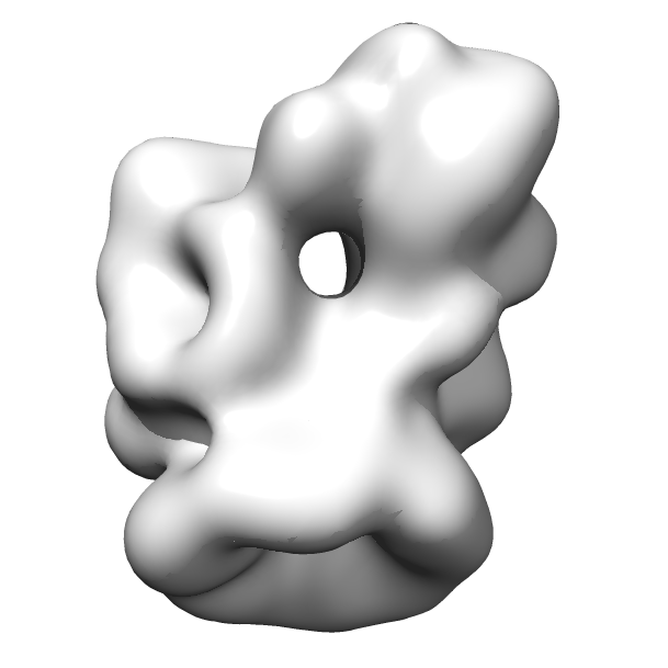 emd_5626.png emd_5626.png | 79.4 KB | ||
| Archive directory |  http://ftp.pdbj.org/pub/emdb/structures/EMD-5626ftp://ftp.pdbj.org/pub/emdb/structures/EMD-5626 http://ftp.pdbj.org/pub/emdb/structures/EMD-5626ftp://ftp.pdbj.org/pub/emdb/structures/EMD-5626 | HTTPS FTP |
-Related structure data


-Links
| EMDB pages | EMDB (EBI/PDBe) / EMDataResource |
|---|
-Map
| File | Download / File: emd_5626.map.gz / Format: CCP4 / Size: 12.6 MB / Type: IMAGE STORED AS FLOATING POINT NUMBER (4 BYTES) | ||||||||||||||||||||||||||||||||||||||||||||||||||||||||||||||||||||
|---|---|---|---|---|---|---|---|---|---|---|---|---|---|---|---|---|---|---|---|---|---|---|---|---|---|---|---|---|---|---|---|---|---|---|---|---|---|---|---|---|---|---|---|---|---|---|---|---|---|---|---|---|---|---|---|---|---|---|---|---|---|---|---|---|---|---|---|---|---|
| Annotation | 3D Cryo-negative EM structure of SWR1 | ||||||||||||||||||||||||||||||||||||||||||||||||||||||||||||||||||||
| Voxel size | X=Y=Z: 3.45 Å | ||||||||||||||||||||||||||||||||||||||||||||||||||||||||||||||||||||
| Density |
| ||||||||||||||||||||||||||||||||||||||||||||||||||||||||||||||||||||
| Symmetry | Space group: 1 | ||||||||||||||||||||||||||||||||||||||||||||||||||||||||||||||||||||
| Details | EMDB XML:
CCP4 map header:
| ||||||||||||||||||||||||||||||||||||||||||||||||||||||||||||||||||||
-Supplemental data
- Sample components
-Entire : ATP-dependent chromatin remodeling complex SWR1
| Entire | Name: ATP-dependent chromatin remodeling complex SWR1 |
|---|---|
| Components |
|
-Supramolecule #1000: ATP-dependent chromatin remodeling complex SWR1
| Supramolecule | Name: ATP-dependent chromatin remodeling complex SWR1 / type: sample / ID: 1000 / Number unique components: 14 |
|---|---|
| Molecular weight | Theoretical: 1.0 MDa |
-Macromolecule #1: SWR1
| Macromolecule | Name: SWR1 / type: protein_or_peptide / ID: 1 / Recombinant expression: No / Database: NCBI |
|---|---|
| Source (natural) | Organism: Saccharomyces cerevisiae (brewer's yeast) / Strain: W1588C-4C / synonym: Baker's yeast |
| Molecular weight | Theoretical: 1.0 MDa |
-Experimental details
-Structure determination
| Method | negative staining, cryo EM |
|---|---|
 Processing Processing | single particle reconstruction |
| Aggregation state | particle |
-Sample preparation
| Buffer | pH: 7.6 Details: 25 mM HEPES-KOH, 1 mM EDTA, 2 mM MgCl2, 0.01% NP-40, 1 mM DTT, 100 mM KCl |
|---|---|
| Staining | Type: NEGATIVE Details: Sample was adsorbed for 15-30 minutes at 4 degrees C. Grid was rinsed with drops of stain (2% uranyl formate) and then a second layer of thin carbon was floated onto the grid. After ...Details: Sample was adsorbed for 15-30 minutes at 4 degrees C. Grid was rinsed with drops of stain (2% uranyl formate) and then a second layer of thin carbon was floated onto the grid. After blotting, the grid was frozen in liquid nitrogen. |
| Grid | Details: Cu 200-mesh Quantifoil grids with thin carbon support, glow discharged in air |
| Vitrification | Cryogen name: NITROGEN / Instrument: OTHER / Method: Cryo-negative staining with manual freezing |
- Electron microscopy
| Microscope | FEI TECNAI F20 |
|---|---|
| Electron beam | Acceleration voltage: 120 kV / Electron source: FIELD EMISSION GUN |
| Electron optics | Calibrated magnification: 87000 / Illumination mode: FLOOD BEAM / Imaging mode: BRIGHT FIELDBright-field microscopy / Cs: 2.0 mm / Nominal defocus max: 2.0 µm / Nominal defocus min: 0.5 µm / Nominal magnification: 62000 |
| Sample stage | Specimen holder model: GATAN LIQUID NITROGEN |
| Temperature | Average: 100 K |
| Date | May 20, 2012 |
| Image recording | Digitization - Sampling interval: 15 µm / Number real images: 300 / Average electron dose: 20 e/Å2 / Camera length: 15 |
| Experimental equipment |  Model: Tecnai F20 / Image courtesy: FEI Company |
-Image processing
| CTF correction | Details: CTFTILT (Grigorieff) for initial model; EMAN2 for projection-matching refinement |
|---|---|
| Final angle assignment | Details: SPIDER |
| Final reconstruction | Algorithm: OTHER / Resolution.type: BY AUTHOR / Resolution: 28.0 Å / Resolution method: FSC 0.5 CUT-OFF / Software - Name: EMAN1, EMAN2, IMAGIC, SPIDER / Number images used: 18000 |
| Details | In order to extract the molecular images from the micrographs, particles were windowed out in one set of micrographs (-45 degrees) using the Boxer interface in EMAN1. Custom-built SPIDER scripts were used to calculate alignment parameters between the +45 degree and -45 degree micrographs and to extract the tilt mates in the +45 degree micrographs. The Contrast Transfer Function (CTF) was estimated and corrected for using the program CTFTILT and the SPIDER command TF CT. Single particles were binned by 2, resulting in a pixel size of 3.3 Angstrom. The +45 degree and -45 degree datasets were combined into a stack of ~32,000 particles and 2D alignment and classification were performed in IMAGIC. Initial models were computed from classes containing 100-200 members using the Orthogonal Tilt Reconstruction approach as described. For projection-matching refinement, the OTR models were initially refined against 2D class averages of cryo-negative data. To generate the class averages, particles were extracted from the micrographs as described above and CTF estimation and phase flipping were performed using the EMAN2 workflow. The particles were then binned by 2, resulting in a pixel size of 3.45 Angstrom. Approximately 32,000 particles were subjected to reference-free 2D alignment and classification in IMAGIC. In order to minimize heterogeneity, classes were generated with relatively few (15-20) particles. The OTR models were filtered to 80 Angstrom resolution and 15-23 iterations of projection matching refinement were performed using angular steps of 250, 200, 150, 100, and 80-50 against 2D class averages in SPIDER using the AP SH and BP 32F commands. To minimize noise in the reconstructions, a threshold mask calculated for 500% to 150%of the theoretical molecular weight of the sample (1.0 MDa) was applied. The mask was gradually tightened throughout refinement and its filtration was determined by the resolution of the 3D map, computed according to the 0.5 FSC criterion. Refinement results were stable after 15 iterations, and the resolutions of the 3D maps were 50-60 Angstrom. The resulting 3D maps (without additional filtration) were then similarly refined against single cryo-negative particles. For this step, 15 iterations of projection-matching refinement were carried out at angular steps of 250, 210, 180, 150, 130, 110 and 100-40. Threshold masks computed for 500% to 100% of the MW were also utilized. |
