 Movie
Movie Controller


+ Open data

- Basic information
| Entry | Database: EMDB / ID: EMD-5600 | |||||||||
|---|---|---|---|---|---|---|---|---|---|---|
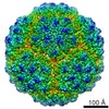
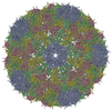
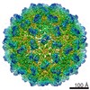
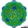
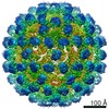
| Title | Penicillium Chrysogenum Virus (PcV) capsid structure | |||||||||
 Map data Map data | Reconstruction of Penicillium Chrysogenum Virus at near-atomic resolution | |||||||||
 Sample Sample |
| |||||||||
 Keywords Keywords |  double-stranded RNA / fungal virus / viral structure / T=1 virus / duplicated helical fold double-stranded RNA / fungal virus / viral structure / T=1 virus / duplicated helical fold | |||||||||
| Function / homology | T=1 icosahedral viral capsid / Capsid protein Function and homology information Function and homology information | |||||||||
| Biological species |  Penicillium chrysogenum virus Penicillium chrysogenum virus | |||||||||
| Method | single particle reconstruction / cryo EM / Resolution: 4.1 Å | |||||||||
 Authors Authors | Luque D / Gomez-Blanco J / Garriga D / Brilot A / Gonzalez JM / Havens WH / Carrascosa JL / Trus BL / Verdaguer N / Grigorieff N ...Luque D / Gomez-Blanco J / Garriga D / Brilot A / Gonzalez JM / Havens WH / Carrascosa JL / Trus BL / Verdaguer N / Grigorieff N / Ghabrial SA / Caston JR | |||||||||
 Citation Citation | Journal: Proc Natl Acad Sci U S A / Year: 2014 Title: Cryo-EM near-atomic structure of a dsRNA fungal virus shows ancient structural motifs preserved in the dsRNA viral lineage. Authors: Daniel Luque / Josué Gómez-Blanco / Damiá Garriga / Axel F Brilot / José M González / Wendy M Havens / José L Carrascosa / Benes L Trus / Nuria Verdaguer / Said A Ghabrial / José R Castón /   Abstract: Viruses evolve so rapidly that sequence-based comparison is not suitable for detecting relatedness among distant viruses. Structure-based comparisons suggest that evolution led to a small number of ...Viruses evolve so rapidly that sequence-based comparison is not suitable for detecting relatedness among distant viruses. Structure-based comparisons suggest that evolution led to a small number of viral classes or lineages that can be grouped by capsid protein (CP) folds. Here, we report that the CP structure of the fungal dsRNA Penicillium chrysogenum virus (PcV) shows the progenitor fold of the dsRNA virus lineage and suggests a relationship between lineages. Cryo-EM structure at near-atomic resolution showed that the 982-aa PcV CP is formed by a repeated α-helical core, indicative of gene duplication despite lack of sequence similarity between the two halves. Superimposition of secondary structure elements identified a single "hotspot" at which variation is introduced by insertion of peptide segments. Structural comparison of PcV and other distantly related dsRNA viruses detected preferential insertion sites at which the complexity of the conserved α-helical core, made up of ancestral structural motifs that have acted as a skeleton, might have increased, leading to evolution of the highly varied current structures. Analyses of structural motifs only apparent after systematic structural comparisons indicated that the hallmark fold preserved in the dsRNA virus lineage shares a long (spinal) α-helix tangential to the capsid surface with the head-tailed phage and herpesvirus viral lineage. | |||||||||
| History |
|
- Structure visualization
| Movie |
Movie viewer |
|---|---|
| Structure viewer | EM map: SurfViewMolmilJmol/JSmol |
| Supplemental images |

- Downloads & links
-EMDB archive
| Map data | emd_5600.map.gz | 219.7 MB | EMDB map data format | |
|---|---|---|---|---|
| Header (meta data) | emd-5600-v30.xmlemd-5600.xml | 10.4 KB 10.4 KB | Display Display | EMDB header |
| Images | 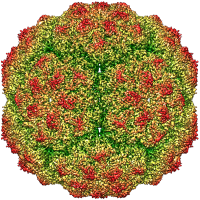 emd_5600.png emd_5600.png | 343.8 KB | ||
| Archive directory |  http://ftp.pdbj.org/pub/emdb/structures/EMD-5600ftp://ftp.pdbj.org/pub/emdb/structures/EMD-5600 http://ftp.pdbj.org/pub/emdb/structures/EMD-5600ftp://ftp.pdbj.org/pub/emdb/structures/EMD-5600 | HTTPS FTP |
-Related structure data
| Related structure data |  3j3iMC M: atomic model generated by this map C: citing same article ( |
|---|---|
| Similar structure data |
-Links
| EMDB pages | EMDB (EBI/PDBe) / EMDataResource |
|---|---|
| Related items in Molecule of the Month |

-Map
| File | Download / File: emd_5600.map.gz / Format: CCP4 / Size: 231.3 MB / Type: IMAGE STORED AS FLOATING POINT NUMBER (4 BYTES) | ||||||||||||||||||||||||||||||||||||||||||||||||||||||||||||||||||||
|---|---|---|---|---|---|---|---|---|---|---|---|---|---|---|---|---|---|---|---|---|---|---|---|---|---|---|---|---|---|---|---|---|---|---|---|---|---|---|---|---|---|---|---|---|---|---|---|---|---|---|---|---|---|---|---|---|---|---|---|---|---|---|---|---|---|---|---|---|---|
| Annotation | Reconstruction of Penicillium Chrysogenum Virus at near-atomic resolution | ||||||||||||||||||||||||||||||||||||||||||||||||||||||||||||||||||||
| Voxel size | X=Y=Z: 1.23 Å | ||||||||||||||||||||||||||||||||||||||||||||||||||||||||||||||||||||
| Density |
| ||||||||||||||||||||||||||||||||||||||||||||||||||||||||||||||||||||
| Symmetry | Space group: 1 | ||||||||||||||||||||||||||||||||||||||||||||||||||||||||||||||||||||
| Details | EMDB XML:
CCP4 map header:
| ||||||||||||||||||||||||||||||||||||||||||||||||||||||||||||||||||||
-Supplemental data
- Sample components
-Entire : Penicillium Chrysogenum Virus
| Entire | Name: Penicillium Chrysogenum Virus |
|---|---|
| Components |
|
-Supramolecule #1000: Penicillium Chrysogenum Virus
| Supramolecule | Name: Penicillium Chrysogenum Virus / type: sample / ID: 1000 / Oligomeric state: icosahedral / Number unique components: 1 |
|---|---|
| Molecular weight | Theoretical: 6.5 MDa |
-Supramolecule #1: Penicillium chrysogenum virus
| Supramolecule | Name: Penicillium chrysogenum virus / type: virus / ID: 1 / NCBI-ID: 158372 / Sci species name: Penicillium chrysogenum virus / Sci species strain: from ATCC 9480 host / Database: NCBI / Virus type: VIRION / Virus isolate: STRAIN / Virus enveloped: No / Virus empty: No |
|---|---|
| Host (natural) | Organism:  Penicillium chrysogenum (fungus) / Strain: ATCC 9480 / synonym: FUNGI Penicillium chrysogenum (fungus) / Strain: ATCC 9480 / synonym: FUNGI |
| Molecular weight | Theoretical: 6.5 MDa |
| Virus shell | Shell ID: 1 / Name: CP / Diameter: 400 Å / T number (triangulation number): 1 |
-Experimental details
-Structure determination
| Method | cryo EM |
|---|---|
 Processing Processing | single particle reconstruction |
| Aggregation state | particle |
-Sample preparation
| Buffer | pH: 7.8 / Details: 50 mM Tris-HCl , 150 mM NaCl, 5 mM EDTA |
|---|---|
| Grid | Details: C flat CF 1/2 4C grids |
| Vitrification | Cryogen name: ETHANE / Chamber humidity: 80 % / Instrument: FEI VITROBOT MARK II Method: Samples were applied to grids, blotted 7 seconds, and plunged into liquid ethane. |
- Electron microscopy
| Microscope | FEI TECNAI F30 |
|---|---|
| Electron beam | Acceleration voltage: 300 kV / Electron source: FIELD EMISSION GUN |
| Electron optics | Calibrated magnification: 56910 / Illumination mode: FLOOD BEAM / Imaging mode: BRIGHT FIELDBright-field microscopy / Cs: 2.0 mm / Nominal defocus max: 3.8 µm / Nominal defocus min: 1.1 µm / Nominal magnification: 58333 |
| Sample stage | Specimen holder model: GATAN LIQUID NITROGEN |
| Date | Feb 23, 2012 |
| Image recording | Category: FILM / Film or detector model: KODAK SO-163 FILM / Digitization - Scanner: ZEISS SCAI / Digitization - Sampling interval: 7 µm / Number real images: 650 / Average electron dose: 25 e/Å2 / Bits/pixel: 8 |
| Experimental equipment |  Model: Tecnai F30 / Image courtesy: FEI Company |
-Image processing
| CTF correction | Details: Phase flipping & amplitude |
|---|---|
| Final reconstruction | Algorithm: OTHER / Resolution.type: BY AUTHOR / Resolution: 4.1 Å / Resolution method: FSC 0.143 CUT-OFF / Software - Name: Xmipp / Number images used: 27566 |
