Movie
Movie Controller


+ Open data

- Basic information
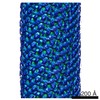
| Entry | Database: EMDB / ID: EMD-5582 | |||||||||
|---|---|---|---|---|---|---|---|---|---|---|
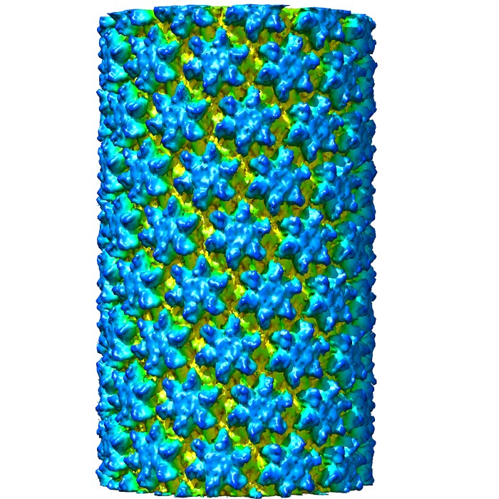
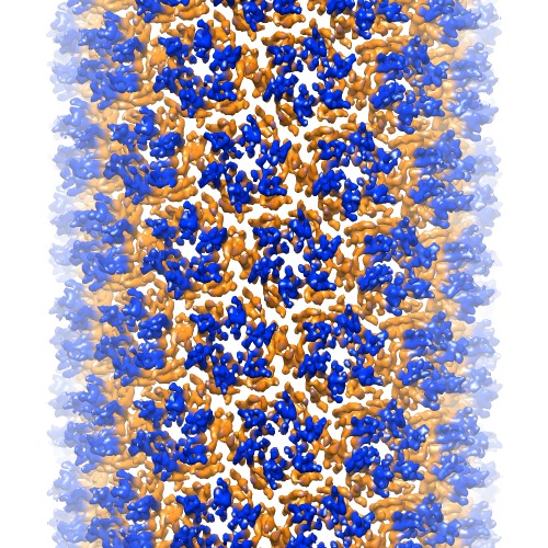
| Title | Cryo-EM structure of HIV-1 capsid assembly | |||||||||
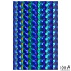
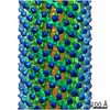
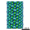
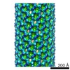
 Map data Map data | Real space helical reconstruction of HIV-1 capsid assembly | |||||||||
 Sample Sample |
| |||||||||
 Keywords Keywords | HIV-1 capsid / core / tubular assembly /  hexamer hexamer | |||||||||
| Function / homology |  Function and homology information Function and homology informationviral budding via host ESCRT complex / viral nucleocapsid / host cell cytoplasm / structural molecule activity / virion membrane / RNA binding / zinc ion binding / identical protein binding / cytoplasmSimilarity search - Function | |||||||||
| Biological species |   Human immunodeficiency virus 1 Human immunodeficiency virus 1 | |||||||||
| Method | helical reconstruction / cryo EM / Resolution: 8.6 Å | |||||||||
 Authors Authors | Zhao G / Perilla JR / Yufenyuy E / Meng X / Chen B / Ning J / Ahn J / Gronenborn AM / Schulten K / Aiken C / Zhang P | |||||||||
 Citation Citation | Journal: Nature / Year: 2013 Title: Mature HIV-1 capsid structure by cryo-electron microscopy and all-atom molecular dynamics. Authors: Gongpu Zhao / Juan R Perilla / Ernest L Yufenyuy / Xin Meng / Bo Chen / Jiying Ning / Jinwoo Ahn / Angela M Gronenborn / Klaus Schulten / Christopher Aiken / Peijun Zhang /  Abstract: Retroviral capsid proteins are conserved structurally but assemble into different morphologies. The mature human immunodeficiency virus-1 (HIV-1) capsid is best described by a 'fullerene cone' model, ...Retroviral capsid proteins are conserved structurally but assemble into different morphologies. The mature human immunodeficiency virus-1 (HIV-1) capsid is best described by a 'fullerene cone' model, in which hexamers of the capsid protein are linked to form a hexagonal surface lattice that is closed by incorporating 12 capsid-protein pentamers. HIV-1 capsid protein contains an amino-terminal domain (NTD) comprising seven α-helices and a β-hairpin, a carboxy-terminal domain (CTD) comprising four α-helices, and a flexible linker with a 310-helix connecting the two structural domains. Structures of the capsid-protein assembly units have been determined by X-ray crystallography; however, structural information regarding the assembled capsid and the contacts between the assembly units is incomplete. Here we report the cryo-electron microscopy structure of a tubular HIV-1 capsid-protein assembly at 8 Å resolution and the three-dimensional structure of a native HIV-1 core by cryo-electron tomography. The structure of the tubular assembly shows, at the three-fold interface, a three-helix bundle with critical hydrophobic interactions. Mutagenesis studies confirm that hydrophobic residues in the centre of the three-helix bundle are crucial for capsid assembly and stability, and for viral infectivity. The cryo-electron-microscopy structures enable modelling by large-scale molecular dynamics simulation, resulting in all-atom models for the hexamer-of-hexamer and pentamer-of-hexamer elements as well as for the entire capsid. Incorporation of pentamers results in closer trimer contacts and induces acute surface curvature. The complete atomic HIV-1 capsid model provides a platform for further studies of capsid function and for targeted pharmacological intervention. | |||||||||
| History |
|
- Structure visualization
| Movie |
Movie viewer |
|---|---|
| Structure viewer | EM map: SurfViewMolmilJmol/JSmol |
| Supplemental images |
- Downloads & links
-EMDB archive
| Map data | emd_5582.map.gz | 932.4 MB | EMDB map data format | |
|---|---|---|---|---|
| Header (meta data) | emd-5582-v30.xmlemd-5582.xml | 12 KB 12 KB | Display Display | EMDB header |
| Images |  emd_5582_1.jpgemd_5582_2.tif emd_5582_1.jpgemd_5582_2.tif | 161.7 KB 583.7 KB | ||
| Archive directory |  http://ftp.pdbj.org/pub/emdb/structures/EMD-5582ftp://ftp.pdbj.org/pub/emdb/structures/EMD-5582 http://ftp.pdbj.org/pub/emdb/structures/EMD-5582ftp://ftp.pdbj.org/pub/emdb/structures/EMD-5582 | HTTPS FTP |
-Related structure data
| Related structure data |  3j34MC  3j4fMC 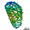 5639C  3j3qC  3j3yC M: atomic model generated by this map C: citing same article ( |
|---|---|
| Similar structure data |
-Links
| EMDB pages | EMDB (EBI/PDBe) / EMDataResource |
|---|---|
| Related items in Molecule of the Month |


-Map
| File | Download / File: emd_5582.map.gz / Format: CCP4 / Size: 1.1 GB / Type: IMAGE STORED AS FLOATING POINT NUMBER (4 BYTES) | ||||||||||||||||||||||||||||||||||||||||||||||||||||||||||||||||||||
|---|---|---|---|---|---|---|---|---|---|---|---|---|---|---|---|---|---|---|---|---|---|---|---|---|---|---|---|---|---|---|---|---|---|---|---|---|---|---|---|---|---|---|---|---|---|---|---|---|---|---|---|---|---|---|---|---|---|---|---|---|---|---|---|---|---|---|---|---|---|
| Annotation | Real space helical reconstruction of HIV-1 capsid assembly | ||||||||||||||||||||||||||||||||||||||||||||||||||||||||||||||||||||
| Voxel size | X=Y=Z: 1.09 Å | ||||||||||||||||||||||||||||||||||||||||||||||||||||||||||||||||||||
| Density |
| ||||||||||||||||||||||||||||||||||||||||||||||||||||||||||||||||||||
| Symmetry | Space group: 1 | ||||||||||||||||||||||||||||||||||||||||||||||||||||||||||||||||||||
| Details | EMDB XML:
CCP4 map header:
| ||||||||||||||||||||||||||||||||||||||||||||||||||||||||||||||||||||
-Supplemental data
- Sample components
-Entire : HIV-1 CA A92E
| Entire | Name: HIV-1 CA A92E |
|---|---|
| Components |
|
-Supramolecule #1000: HIV-1 CA A92E
| Supramolecule | Name: HIV-1 CA A92E / type: sample / ID: 1000 / Oligomeric state: helical assembly of hexamers / Number unique components: 6 |
|---|---|
| Molecular weight | Experimental: 25 KDa / Theoretical: 25 KDa |
-Macromolecule #1: HIV-1 capsid protein
| Macromolecule | Name: HIV-1 capsid protein / type: protein_or_peptide / ID: 1 / Name.synonym: CA / Oligomeric state: hexamer / Recombinant expression: Yes |
|---|---|
| Source (natural) | Organism: Human immunodeficiency virus 1 / synonym: HIV-1 |
| Molecular weight | Experimental: 25 KDa / Theoretical: 25 KDa |
| Recombinant expression | Organism:  Escherichia coli (E. coli) / Recombinant strain: Rosetta 2 (DE3) / Recombinant plasmid: pET21 Escherichia coli (E. coli) / Recombinant strain: Rosetta 2 (DE3) / Recombinant plasmid: pET21 |
-Experimental details
-Structure determination
| Method | cryo EM |
|---|---|
 Processing Processing | helical reconstruction |
| Aggregation state | filament |
-Sample preparation
| Concentration | 2 mg/mL |
|---|---|
| Buffer | pH: 8 / Details: 1 M NaCl, 50 mM Tris-HCl |
| Grid | Details: 200 mesh quantifoil R2/1 copper grid |
| Vitrification | Cryogen name: ETHANE / Chamber humidity: 80 % / Chamber temperature: 90 K / Instrument: HOMEMADE PLUNGER Method: With 2.5 uL sample on carbon side, add 3 uL dilution buffer (100 mM NaCl, 50 mM Tris, pH 8.0) to back side. Blot 3-5 seconds from back side. |
- Electron microscopy
| Microscope | FEI POLARA 300 |
|---|---|
| Electron beam | Acceleration voltage: 200 kV / Electron source: FIELD EMISSION GUN |
| Electron optics | Calibrated magnification: 58257 / Illumination mode: FLOOD BEAM / Imaging mode: BRIGHT FIELDBright-field microscopy / Cs: 2 mm / Nominal defocus max: 3.5 µm / Nominal defocus min: 1.0 µm / Nominal magnification: 59000 |
| Sample stage | Specimen holder model: SIDE ENTRY, EUCENTRIC |
| Temperature | Min: 80 K / Max: 85 K / Average: 82 K |
| Date | Dec 11, 2010 |
| Image recording | Category: FILM / Film or detector model: KODAK SO-163 FILM / Digitization - Scanner: NIKON SUPER COOLSCAN 9000 / Digitization - Sampling interval: 6.35 µm / Number real images: 27 / Average electron dose: 15 e/Å2 / Bits/pixel: 16 |
| Experimental equipment |  Model: Tecnai Polara / Image courtesy: FEI Company |
-Image processing
| CTF correction | Details: each filament |
|---|---|
| Final reconstruction | Applied symmetry - Helical parameters - Δz: 7.247 Å Applied symmetry - Helical parameters - Δ&Phi: 31.13 ° Resolution.type: BY AUTHOR / Resolution: 8.6 Å / Resolution method: FSC 0.5 CUT-OFF / Software - Name: Frealign |
| Details | The segments were aligned and reconstructed using Frealign. Twofold symmetry was imposed using IHRSR++. |
-Atomic model buiding 1
| Initial model | PDB ID: |
|---|---|
| Software | Name: MDFF |
| Refinement | Space: REAL / Protocol: FLEXIBLE FIT |
| Output model | PDB-3j34: PDB-3j4f: |