 Movie
Movie Controller

[English] 日本語
 Yorodumi
- EMDB-5524: A pseudoatomic model of the COPII cage obtained from cryo-electro... -
+ Open data

- Basic information
| Entry | Database: EMDB / ID: EMD-5524 | |||||||||
|---|---|---|---|---|---|---|---|---|---|---|
| Title | A pseudoatomic model of the COPII cage obtained from cryo-electron microscopy and mass spectrometry | |||||||||
 Map data Map data | Reconstruction of the Sec13/31 COPII cage | |||||||||
 Sample Sample |
| |||||||||
 Keywords Keywords | Sec13 Sec31 COPII | |||||||||
| Function / homology | COPII vesicle coat /  intracellular protein transport / WD40 repeat intracellular protein transport / WD40 repeat Function and homology information Function and homology information | |||||||||
| Biological species |  Homo sapiens (human) Homo sapiens (human) | |||||||||
| Method | single particle reconstruction / cryo EM / Resolution: 12.0 Å | |||||||||
 Authors Authors | Noble AJ / Zhang Q / O'Donnell J / Hariri H / Bhattacharya N / Marshall AG / Stagg SM | |||||||||
 Citation Citation | Journal: Nat Struct Mol Biol / Year: 2013 Title: A pseudoatomic model of the COPII cage obtained from cryo-electron microscopy and mass spectrometry. Authors: Alex J Noble / Qian Zhang / Jason O'Donnell / Hanaa Hariri / Nilakshee Bhattacharya / Alan G Marshall / Scott M Stagg /  Abstract: COPII vesicles transport proteins from the endoplasmic reticulum to the Golgi apparatus. Previous COPII-cage cryo-EM structures lacked the resolution necessary to determine the residues of Sec13 and ...COPII vesicles transport proteins from the endoplasmic reticulum to the Golgi apparatus. Previous COPII-cage cryo-EM structures lacked the resolution necessary to determine the residues of Sec13 and Sec31 that mediate assembly and flexibility of the COPII cage. Here we present a 12-Å structure of the human COPII cage, where the tertiary structure of Sec13 and Sec31 is clearly identifiable. We employ this structure and a homology model of the Sec13-Sec31 complex to create a reliable pseudoatomic model of the COPII cage. We combined this model with hydrogen/deuterium-exchange MS analysis to characterize four distinct contact regions at the vertices of the COPII cage. Furthermore, we found that the two-fold symmetry of the Sec31 dimeric region in Sec13-Sec31 is broken upon cage formation and that the resulting hinge is essential to form the proper edge geometry in COPII cages. | |||||||||
| History |
|
- Structure visualization
| Movie |
Movie viewer |
|---|---|
| Structure viewer | EM map: SurfViewMolmilJmol/JSmol |
| Supplemental images |

- Downloads & links
-EMDB archive
| Map data | emd_5524.map.gz | 81.4 MB | EMDB map data format | |
|---|---|---|---|---|
| Header (meta data) | emd-5524-v30.xmlemd-5524.xml | 11.6 KB 11.6 KB | Display Display | EMDB header |
| Images | 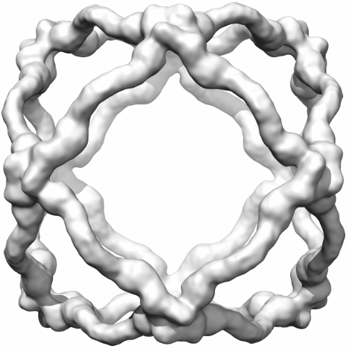 emd_5524_1.png emd_5524_1.png | 162.8 KB | ||
| Archive directory |  http://ftp.pdbj.org/pub/emdb/structures/EMD-5524ftp://ftp.pdbj.org/pub/emdb/structures/EMD-5524 http://ftp.pdbj.org/pub/emdb/structures/EMD-5524ftp://ftp.pdbj.org/pub/emdb/structures/EMD-5524 | HTTPS FTP |
-Related structure data
| Similar structure data |
|---|


-Links
| EMDB pages | EMDB (EBI/PDBe) / EMDataResource |
|---|
-Map
| File | Download / File: emd_5524.map.gz / Format: CCP4 / Size: 89 MB / Type: IMAGE STORED AS FLOATING POINT NUMBER (4 BYTES) | ||||||||||||||||||||||||||||||||||||||||||||||||||||||||||||||||||||
|---|---|---|---|---|---|---|---|---|---|---|---|---|---|---|---|---|---|---|---|---|---|---|---|---|---|---|---|---|---|---|---|---|---|---|---|---|---|---|---|---|---|---|---|---|---|---|---|---|---|---|---|---|---|---|---|---|---|---|---|---|---|---|---|---|---|---|---|---|---|
| Annotation | Reconstruction of the Sec13/31 COPII cage | ||||||||||||||||||||||||||||||||||||||||||||||||||||||||||||||||||||
| Voxel size | X=Y=Z: 2.78 Å | ||||||||||||||||||||||||||||||||||||||||||||||||||||||||||||||||||||
| Density |
| ||||||||||||||||||||||||||||||||||||||||||||||||||||||||||||||||||||
| Symmetry | Space group: 1 | ||||||||||||||||||||||||||||||||||||||||||||||||||||||||||||||||||||
| Details | EMDB XML:
CCP4 map header:
| ||||||||||||||||||||||||||||||||||||||||||||||||||||||||||||||||||||
-Supplemental data
- Sample components
-Entire : Oligomeric assembly of Sec13/31
| Entire | Name: Oligomeric assembly of Sec13/31 |
|---|---|
| Components |
|
-Supramolecule #1000: Oligomeric assembly of Sec13/31
| Supramolecule | Name: Oligomeric assembly of Sec13/31 / type: sample / ID: 1000 Oligomeric state: Each Sec13/31 heterotetramer consists of two Sec13s and two Sec31s. Number unique components: 2 |
|---|---|
| Molecular weight | Theoretical: 8.08 MDa |
-Macromolecule #1: Sec13R
| Macromolecule | Name: Sec13R / type: protein_or_peptide / ID: 1 / Name.synonym: SEC13 / Number of copies: 2 / Oligomeric state: Heterotetramer / Recombinant expression: Yes |
|---|---|
| Source (natural) | Organism: Homo sapiens (human) / synonym: Human / Location in cell: cytosol |
| Molecular weight | Theoretical: 35.4 KDa |
| Recombinant expression | Organism:  Trichoplusia ni (cabbage looper) / Recombinant plasmid: pFastBac Dual Trichoplusia ni (cabbage looper) / Recombinant plasmid: pFastBac Dual |
| Sequence | GO: intracellular protein transport / InterPro: WD40 repeat |
-Macromolecule #2: Sec31L1
| Macromolecule | Name: Sec31L1 / type: protein_or_peptide / ID: 2 / Name.synonym: SEC31 / Number of copies: 2 / Oligomeric state: Heterotetramer / Recombinant expression: Yes |
|---|---|
| Source (natural) | Organism: Homo sapiens (human) / synonym: Human / Location in cell: cytosol |
| Molecular weight | Theoretical: 133 KDa |
| Recombinant expression | Organism: Trichoplusia ni (cabbage looper) / Recombinant plasmid: pFastBAC Dual |
| Sequence | GO: COPII vesicle coat / InterPro: WD40 repeat |
-Experimental details
-Structure determination
| Method | cryo EM |
|---|---|
 Processing Processing | single particle reconstruction |
| Aggregation state | particle |
-Sample preparation
| Concentration | 3 mg/mL |
|---|---|
| Buffer | pH: 7.5 Details: 20 mM Tris-Cl, pH 7.5, 700 mM KOAc, 1 mM MgOAc, 10 mM DTT |
| Grid | Details: Quantifoil R2/1 grids plasma cleaned for 8 s |
| Vitrification | Cryogen name: ETHANE / Chamber humidity: 100 % / Chamber temperature: 93 K / Instrument: FEI VITROBOT MARK IV Method: Add 3ul of sample onto Quantifoil R2/1 grids and plasma clean for 8s using a Gatan Solarus plasma cleaner. |
- Electron microscopy
| Microscope | FEI TITAN KRIOS |
|---|---|
| Electron beam | Acceleration voltage: 120 kV / Electron source: FIELD EMISSION GUN |
| Electron optics | Illumination mode: FLOOD BEAM / Imaging mode: BRIGHT FIELDBright-field microscopy / Cs: 2.7 mm / Nominal defocus max: 3.5 µm / Nominal defocus min: 1.5 µm / Nominal magnification: 37000 |
| Sample stage | Specimen holder: liquid nitrogen cooled / Specimen holder model: FEI TITAN KRIOS AUTOGRID HOLDER |
| Temperature | Average: 94 K |
| Alignment procedure | Legacy - Astigmatism: Objective lens astigmatism was corrected at 120,000 times magnification |
| Date | Jun 24, 2011 |
| Image recording | Category: CCD / Film or detector model: GATAN ULTRASCAN 4000 (4k x 4k) / Number real images: 5052 / Average electron dose: 15 e/Å2 |
| Experimental equipment |  Model: Titan Krios / Image courtesy: FEI Company |
-Image processing
| CTF correction | Details: phase flip of each particle |
|---|---|
| Final two d classification | Number classes: 756 |
| Final reconstruction | Algorithm: OTHER / Resolution.type: BY AUTHOR / Resolution: 12.0 Å / Resolution method: FSC 0.143 CUT-OFF / Software - Name: EMAN, Spider Details: Later refinements used proc3d's automask2 option to mask out densities internal to the Sec13/31 cage. Number images used: 23404 |
| Details | The particles were selected automatically using template matching. |
