 Movie
Movie Controller

[English] 日本語
 Yorodumi
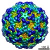
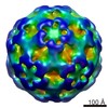
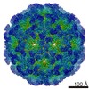
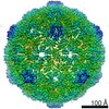
- EMDB-5428: Cryo-EM reconstruction of Nudaurelia capensis omega virus (NwV) c... -
+ Open data

- Basic information
| Entry | Database: EMDB / ID: EMD-5428 | |||||||||
|---|---|---|---|---|---|---|---|---|---|---|
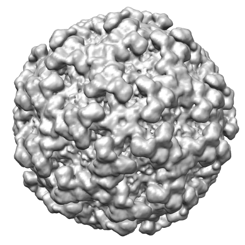
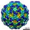
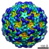
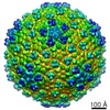
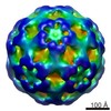
| Title | Cryo-EM reconstruction of Nudaurelia capensis omega virus (NwV) capsid at 4hr time point following maturation initiation | |||||||||
 Map data Map data | Reconstruction of Nudaurelia capensis omega virus (NwV) capsid at 4hr time point following maturation initiation | |||||||||
 Sample Sample |
| |||||||||
 Keywords Keywords | Time-resolved / T=4 /  quasi-equivalence / maturation quasi-equivalence / maturation | |||||||||
| Biological species |  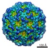 Nudaurelia capensis omega virus Nudaurelia capensis omega virus | |||||||||
| Method | single particle reconstruction / cryo EM / Resolution: 8.3 Å | |||||||||
 Authors Authors | Matsui T / Lander GC / Khayat R / Johnson JE | |||||||||
 Citation Citation | Journal: Proc Natl Acad Sci U S A / Year: 2010 Title: Subunits fold at position-dependent rates during maturation of a eukaryotic RNA virus. Authors: Tsutomu Matsui / Gabriel C Lander / Reza Khayat / John E Johnson /  Abstract: Effective antiviral agents are difficult to develop because of the close relationship between the cell biology of the virus and host. However, viral capsid maturation, the in vivo process where the ...Effective antiviral agents are difficult to develop because of the close relationship between the cell biology of the virus and host. However, viral capsid maturation, the in vivo process where the particle transitions from a noninfectious provirion to an infectious virion, is an ideal process to interrupt because the provirion is usually fragile and the conversion to the virion often involves large conformational changes and autocatalytic chemistry that can be hampered by small molecules. The Nudaurelia capensis omega virus (N omegaV) is one of the few eukaryotic viruses where this process can be investigated in vitro with a variety of biophysical methods, allowing fundamental chemical and structural principles of the maturation to be established. It has a T = 4 quasi-equivalent capsid with a dramatic maturation pathway that includes a particle size reduction of 100 A and an autocatalytic cleavage. Here we use cryo-EM and difference maps, computed at three time points following maturation initiation, to show that regions of N omegaV subunit folding are maturation dependent and occur at rates determined by their quasi-equivalent position in the capsid, explaining the unusual kinetics of the maturation cleavage. This study shows that folding is rapid and peptide chain self-cleavage occurs early for subunits adjacent to 3-fold and 5-fold icosahedral symmetry elements and that folding is slower in regions where molecular switches are required for the formation of the proper interfacial contacts. The results connect viral maturation to the well-studied assembly-dependent folding that occurs in the formation of cellular complexes. | |||||||||
| History |
|
- Structure visualization
| Movie |
 Movie viewer Movie viewer |
|---|---|
| Structure viewer | EM map: SurfViewMolmilJmol/JSmol |
| Supplemental images |
- Downloads & links
-EMDB archive
| Map data | emd_5428.map.gz | 12.5 MB | EMDB map data format | |
|---|---|---|---|---|
| Header (meta data) | emd-5428-v30.xmlemd-5428.xml | 8.8 KB 8.8 KB | Display Display | EMDB header |
| Images |  emd_5428_1.png emd_5428_1.png | 161.1 KB | ||
| Archive directory |  http://ftp.pdbj.org/pub/emdb/structures/EMD-5428ftp://ftp.pdbj.org/pub/emdb/structures/EMD-5428 http://ftp.pdbj.org/pub/emdb/structures/EMD-5428ftp://ftp.pdbj.org/pub/emdb/structures/EMD-5428 | HTTPS FTP |
-Related structure data

-Links
| EMDB pages | EMDB (EBI/PDBe) / EMDataResource |
|---|
-Map
| File | Download / File: emd_5428.map.gz / Format: CCP4 / Size: 29.8 MB / Type: IMAGE STORED AS FLOATING POINT NUMBER (4 BYTES) | ||||||||||||||||||||||||||||||||||||||||||||||||||||||||||||||||||||
|---|---|---|---|---|---|---|---|---|---|---|---|---|---|---|---|---|---|---|---|---|---|---|---|---|---|---|---|---|---|---|---|---|---|---|---|---|---|---|---|---|---|---|---|---|---|---|---|---|---|---|---|---|---|---|---|---|---|---|---|---|---|---|---|---|---|---|---|---|---|
| Annotation | Reconstruction of Nudaurelia capensis omega virus (NwV) capsid at 4hr time point following maturation initiation | ||||||||||||||||||||||||||||||||||||||||||||||||||||||||||||||||||||
| Voxel size | X=Y=Z: 2.768 Å | ||||||||||||||||||||||||||||||||||||||||||||||||||||||||||||||||||||
| Density |
| ||||||||||||||||||||||||||||||||||||||||||||||||||||||||||||||||||||
| Symmetry | Space group: 1 | ||||||||||||||||||||||||||||||||||||||||||||||||||||||||||||||||||||
| Details | EMDB XML:
CCP4 map header:
| ||||||||||||||||||||||||||||||||||||||||||||||||||||||||||||||||||||
-Supplemental data
- Sample components
-Entire : Nudaurelia capensis omega virus (NwV) capsid at 4hr time point fo...
| Entire | Name: Nudaurelia capensis omega virus (NwV) capsid at 4hr time point following maturation initiation |
|---|---|
| Components |
|
-Supramolecule #1000: Nudaurelia capensis omega virus (NwV) capsid at 4hr time point fo...
| Supramolecule | Name: Nudaurelia capensis omega virus (NwV) capsid at 4hr time point following maturation initiation type: sample / ID: 1000 / Oligomeric state: Icosahedral virus / Number unique components: 1 |
|---|
-Supramolecule #1: Nudaurelia capensis omega virus
| Supramolecule | Name: Nudaurelia capensis omega virus / type: virus / ID: 1 / Name.synonym: NwV (N omega V) / NCBI-ID: 12541 / Sci species name: Nudaurelia capensis omega virus / Database: NCBI / Virus type: VIRUS-LIKE PARTICLE / Virus isolate: STRAIN / Virus enveloped: No / Virus empty: No / Syn species name: NwV (N omega V) |
|---|---|
| Host (natural) | Organism:  Lepidoptera (butterflies and moths) / synonym: INVERTEBRATES Lepidoptera (butterflies and moths) / synonym: INVERTEBRATES |
| Virus shell | Shell ID: 1 / Diameter: 400 Å / T number (triangulation number): 4 |
-Experimental details
-Structure determination
| Method | cryo EM |
|---|---|
 Processing Processing | single particle reconstruction |
| Aggregation state | particle |
-Sample preparation
| Concentration | 10 mg/mL |
|---|---|
| Buffer | pH: 5 / Details: 100mM NaOAc, 250mM NaCl |
| Vitrification | Cryogen name: ETHANE / Chamber humidity: 100 % / Chamber temperature: 90 K / Instrument: FEI VITROBOT MARK I / Method: Blot for 5sec before plunging |
- Electron microscopy
| Microscope | FEI TECNAI F20 |
|---|---|
| Electron beam | Acceleration voltage: 120 kV / Electron source: FIELD EMISSION GUN |
| Electron optics | Illumination mode: FLOOD BEAM / Imaging mode: BRIGHT FIELDBright-field microscopy / Nominal defocus max: 3.0 µm / Nominal defocus min: 1.0 µm / Nominal magnification: 80000 |
| Sample stage | Specimen holder model: GATAN LIQUID NITROGEN |
| Date | Feb 13, 2008 |
| Image recording | Number real images: 295 / Average electron dose: 16.5 e/Å2 Details: Data were collected using the Leginon automated electron microscopy package and data processing was performed using the Appion pipeline. |
| Experimental equipment |  Model: Tecnai F20 / Image courtesy: FEI Company |
-Image processing
| CTF correction | Details: Each image |
|---|---|
| Final reconstruction | Algorithm: OTHER / Resolution.type: BY AUTHOR / Resolution: 8.3 Å / Resolution method: FSC 0.5 CUT-OFF / Software - Name: EMAN Details: Data processing was performed using the Appion pipeline. Number images used: 23392 |
