Movie
Movie Controller

[English] 日本語
 Yorodumi
- EMDB-5424: Structure of adeno-associated virus-2 in complex with neutralizin... -
+ Open data

- Basic information
| Entry | Database: EMDB / ID: EMD-5424 | |||||||||
|---|---|---|---|---|---|---|---|---|---|---|
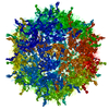
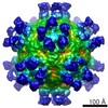
| Title | Structure of adeno-associated virus-2 in complex with neutralizing monoclonal antibody A20 | |||||||||
 Map data Map data | Reconstruction of adeno-associated virus-2 in complex with neutralizing monoclonal antibody A20 | |||||||||
 Sample Sample |
| |||||||||
 Keywords Keywords | Adeno-associated virus / Antibody / A20 / Epitope / Fab / Gene therapy / Monoclonal | |||||||||
| Function / homology |  Function and homology information Function and homology informationpermeabilization of host organelle membrane involved in viral entry into host cell / symbiont entry into host cell via permeabilization of inner membrane / host cell nucleolus / T=1 icosahedral viral capsid / clathrin-dependent endocytosis of virus by host cell / virion attachment to host cell / structural molecule activity Similarity search - Function | |||||||||
| Biological species |  Adeno-associated virus - 2 Adeno-associated virus - 2 | |||||||||
| Method | single particle reconstruction / cryo EM / Resolution: 8.5 Å | |||||||||
 Authors Authors | McCraw DM / O'Donnell JK / Taylor KA / Stagg SM / Chapman MS | |||||||||
 Citation Citation | Journal: Virology Title: Structure of adeno-associated virus-2 in complex with neutralizing monoclonal antibody A20. Authors: Dustin M McCraw / Jason K O'Donnell / Kenneth A Taylor / Scott M Stagg / Michael S Chapman /  Abstract: The use of adeno-associated virus (AAV) as a gene therapy vector is limited by the host neutralizing immune response. The cryo-electron microscopy (EM) structure at 8.5Å resolution is determined for ...The use of adeno-associated virus (AAV) as a gene therapy vector is limited by the host neutralizing immune response. The cryo-electron microscopy (EM) structure at 8.5Å resolution is determined for a complex of AAV-2 with the Fab' fragment of monoclonal antibody (MAb) A20, the most extensively characterized AAV MAb. The binding footprint is determined through fitting the cryo-EM reconstruction with a homology model following sequencing of the variable domain, and provides a structural basis for integrating diverse prior epitope mappings. The footprint extends from the previously implicated plateau to the side of the spike, and into the conserved canyon, covering a larger area than anticipated. Comparison with structures of binding and non-binding serotypes indicates that recognition depends on a combination of subtle serotype-specific features. Separation of the neutralizing epitope from the heparan sulfate cell attachment site encourages attempts to develop immune-resistant vectors that can still bind to target cells. | |||||||||
| History |
|
- Structure visualization
| Movie |
Movie viewer |
|---|---|
| Structure viewer | EM map: SurfViewMolmilJmol/JSmol |
| Supplemental images |

- Downloads & links
-EMDB archive
| Map data | emd_5424.map.gz | 30.7 MB | EMDB map data format | |
|---|---|---|---|---|
| Header (meta data) | emd-5424-v30.xmlemd-5424.xml | 11.2 KB 11.2 KB | Display Display | EMDB header |
| Images | 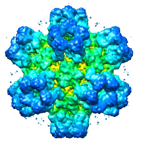 emd_5424_1.jpg emd_5424_1.jpg | 75.9 KB | ||
| Archive directory |  http://ftp.pdbj.org/pub/emdb/structures/EMD-5424ftp://ftp.pdbj.org/pub/emdb/structures/EMD-5424 http://ftp.pdbj.org/pub/emdb/structures/EMD-5424ftp://ftp.pdbj.org/pub/emdb/structures/EMD-5424 | HTTPS FTP |
-Validation report
| Summary document | emd_5424_validation.pdf.gz | 332.6 KB | Display | EMDB validaton report |
|---|---|---|---|---|
| Full document | emd_5424_full_validation.pdf.gz | 332.2 KB | Display | |
| Data in XML | emd_5424_validation.xml.gz | 6.2 KB | Display | |
| Arichive directory | https://ftp.pdbj.org/pub/emdb/validation_reports/EMD-5424ftp://ftp.pdbj.org/pub/emdb/validation_reports/EMD-5424 | HTTPS FTP |
-Related structure data
| Related structure data |  3j1sMC M: atomic model generated by this map C: citing same article ( |
|---|---|
| Similar structure data |
-Links
| EMDB pages | EMDB (EBI/PDBe) / EMDataResource |
|---|---|
| Related items in Molecule of the Month |


-Map
| File | Download / File: emd_5424.map.gz / Format: CCP4 / Size: 51.5 MB / Type: IMAGE STORED AS FLOATING POINT NUMBER (4 BYTES) | ||||||||||||||||||||||||||||||||||||||||||||||||||||||||||||||||||||
|---|---|---|---|---|---|---|---|---|---|---|---|---|---|---|---|---|---|---|---|---|---|---|---|---|---|---|---|---|---|---|---|---|---|---|---|---|---|---|---|---|---|---|---|---|---|---|---|---|---|---|---|---|---|---|---|---|---|---|---|---|---|---|---|---|---|---|---|---|---|
| Annotation | Reconstruction of adeno-associated virus-2 in complex with neutralizing monoclonal antibody A20 | ||||||||||||||||||||||||||||||||||||||||||||||||||||||||||||||||||||
| Voxel size | X=Y=Z: 2.45 Å | ||||||||||||||||||||||||||||||||||||||||||||||||||||||||||||||||||||
| Density |
| ||||||||||||||||||||||||||||||||||||||||||||||||||||||||||||||||||||
| Symmetry | Space group: 1 | ||||||||||||||||||||||||||||||||||||||||||||||||||||||||||||||||||||
| Details | EMDB XML:
CCP4 map header:
| ||||||||||||||||||||||||||||||||||||||||||||||||||||||||||||||||||||
-Supplemental data
- Sample components
-Entire : Adeno-associated virus-2 in complex with neutralizing monoclonal ...
| Entire | Name: Adeno-associated virus-2 in complex with neutralizing monoclonal antibody A20 |
|---|---|
| Components |
|
-Supramolecule #1000: Adeno-associated virus-2 in complex with neutralizing monoclonal ...
| Supramolecule | Name: Adeno-associated virus-2 in complex with neutralizing monoclonal antibody A20 type: sample / ID: 1000 Oligomeric state: 60 A20 Fab's bind to one adeno-associated virus (one adeno-associated virus consists of 60 viral proteins) Number unique components: 2 |
|---|---|
| Molecular weight | Theoretical: 6.9 MDa |
-Supramolecule #1: Adeno-associated virus - 2
| Supramolecule | Name: Adeno-associated virus - 2 / type: virus / ID: 1 / Name.synonym: AAV-2 / NCBI-ID: 10804 / Sci species name: Adeno-associated virus - 2 / Database: NCBI / Virus type: VIRION / Virus isolate: SEROTYPE / Virus enveloped: No / Virus empty: No / Syn species name: AAV-2 |
|---|---|
| Host (natural) | Organism:  Homo sapiens (human) / synonym: VERTEBRATES Homo sapiens (human) / synonym: VERTEBRATES |
| Molecular weight | Theoretical: 3.9 MDa |
| Virus shell | Shell ID: 1 / Diameter: 280 Å / T number (triangulation number): 1 |
-Experimental details
-Structure determination
| Method | cryo EM |
|---|---|
 Processing Processing | single particle reconstruction |
| Aggregation state | particle |
-Sample preparation
| Concentration | 0.14 mg/mL |
|---|---|
| Buffer | pH: 7.2 Details: 100 mM HEPES, 50 mM magnesium chloride, and 5% glycerol |
| Grid | Details: 400 mesh carbon grid with holey carbon support |
| Vitrification | Cryogen name: ETHANE / Chamber humidity: 100 % / Chamber temperature: 90 K / Instrument: FEI VITROBOT MARK IV / Method: Blot for 2.0 seconds before plunging. |
- Electron microscopy
| Microscope | FEI TITAN KRIOS |
|---|---|
| Temperature | Average: 93 K |
| Alignment procedure | Legacy - Astigmatism: Objective lens astigmatism was corrected at 120,000 times magnification Legacy - Electron beam tilt params: 0 |
| Date | Feb 23, 2011 |
| Image recording | Category: CCD / Film or detector model: GATAN ULTRASCAN 1000 (2k x 2k) / Number real images: 1503 / Average electron dose: 15 e/Å2 / Bits/pixel: 16 |
| Electron beam | Acceleration voltage: 120 kV / Electron source:  FIELD EMISSION GUN FIELD EMISSION GUN |
| Electron optics | Calibrated magnification: 39775 / Illumination mode: SPOT SCAN / Imaging mode: BRIGHT FIELD / Cs: 2.7 mm / Nominal defocus max: -4.0 µm / Nominal defocus min: -0.5 µm / Nominal magnification: 37000 |
| Sample stage | Specimen holder: Nitrogen cooled / Specimen holder model: FEI TITAN KRIOS AUTOGRID HOLDER |
| Experimental equipment |  Model: Titan Krios / Image courtesy: FEI Company |
-Image processing
| CTF correction | Details: Whole image |
|---|---|
| Final reconstruction | Algorithm: OTHER / Resolution.type: BY AUTHOR / Resolution: 8.5 Å / Resolution method: FSC 0.5 CUT-OFF / Software - Name: Appion, ACE, EMAN / Number images used: 11898 |
| Final two d classification | Number classes: 304 |
-Atomic model buiding 1
| Initial model | PDB ID: Chain - Chain ID: A |
|---|---|
| Software | Name: RSRef |
| Details | Protocol: Fixed. Anti-bumping restraints from CNS included 1LP3, Constrained icosahedral symmetry |
| Refinement | Space: REAL / Overall B value: 0 Target criteria: Least-squares difference between experimental & model coulombic potential |
| Output model | PDB-3j1s: |