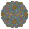
Movie
Movie Controller


+ Open data

- Basic information
| Entry | Database: EMDB / ID: EMD-5406 | |||||||||
|---|---|---|---|---|---|---|---|---|---|---|
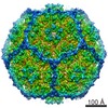
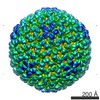
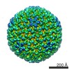
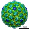
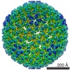
| Title | Icosahedral reconstruction of bacteriophage P2 procapsid | |||||||||
 Map data Map data | Reconstruction of bacteriophage P2 procapsid | |||||||||
 Sample Sample |
| |||||||||
 Keywords Keywords |  bacteriophage / P2 / P4 / capsid / procapsid / assembly / size determination / scaffolding protein / triangulation numbers / T=7 dextro / Sid / gpN bacteriophage / P2 / P4 / capsid / procapsid / assembly / size determination / scaffolding protein / triangulation numbers / T=7 dextro / Sid / gpN | |||||||||
| Biological species |  Escherichia coli (E. coli) Escherichia coli (E. coli) | |||||||||
| Method | single particle reconstruction / cryo EM / Resolution: 8.0 Å | |||||||||
 Authors Authors | Dearborn AD / Laurinmaki P / Chandramouli P / Rodenburg CM / Wang S / Butcher SJ / Dokland T | |||||||||
 Citation Citation | Journal: J Struct Biol / Year: 2012 Title: Structure and size determination of bacteriophage P2 and P4 procapsids: function of size responsiveness mutations. Authors: Altaira D Dearborn / Pasi Laurinmaki / Preethi Chandramouli / Cynthia M Rodenburg / Sifang Wang / Sarah J Butcher / Terje Dokland /  Abstract: Bacteriophage P4 is dependent on structural proteins supplied by a helper phage, P2, to assemble infectious virions. Bacteriophage P2 normally forms an icosahedral capsid with T=7 symmetry from the ...Bacteriophage P4 is dependent on structural proteins supplied by a helper phage, P2, to assemble infectious virions. Bacteriophage P2 normally forms an icosahedral capsid with T=7 symmetry from the gpN capsid protein, the gpO scaffolding protein and the gpQ portal protein. In the presence of P4, however, the same structural proteins are assembled into a smaller capsid with T=4 symmetry. This size determination is effected by the P4-encoded protein Sid, which forms an external scaffold around the small P4 procapsids. Size responsiveness (sir) mutants in gpN fail to assemble small capsids even in the presence of Sid. We have produced large and small procapsids by co-expression of gpN with gpO and Sid, respectively, and applied cryo-electron microscopy and three-dimensional reconstruction methods to visualize these procapsids. gpN has an HK97-like fold and interacts with Sid in an exposed loop where the sir mutations are clustered. The T=7 lattice of P2 has dextro handedness, unlike the laevo lattices of other phages with this fold observed so far. | |||||||||
| History |
|
- Structure visualization
| Movie |
 Movie viewer Movie viewer |
|---|---|
| Structure viewer | EM map: SurfViewMolmilJmol/JSmol |
| Supplemental images |

- Downloads & links
-EMDB archive
| Map data | emd_5406.map.gz | 117.2 MB | EMDB map data format | |
|---|---|---|---|---|
| Header (meta data) | emd-5406-v30.xmlemd-5406.xml | 10.5 KB 10.5 KB | Display Display | EMDB header |
| Images |  emd_5406_1.jpg emd_5406_1.jpg | 118.7 KB | ||
| Archive directory |  http://ftp.pdbj.org/pub/emdb/structures/EMD-5406ftp://ftp.pdbj.org/pub/emdb/structures/EMD-5406 http://ftp.pdbj.org/pub/emdb/structures/EMD-5406ftp://ftp.pdbj.org/pub/emdb/structures/EMD-5406 | HTTPS FTP |
-Related structure data

-Links
| EMDB pages | EMDB (EBI/PDBe) / EMDataResource |
|---|
-Map
| File | Download / File: emd_5406.map.gz / Format: CCP4 / Size: 122.1 MB / Type: IMAGE STORED AS FLOATING POINT NUMBER (4 BYTES) | ||||||||||||||||||||||||||||||||||||||||||||||||||||||||||||||||||||
|---|---|---|---|---|---|---|---|---|---|---|---|---|---|---|---|---|---|---|---|---|---|---|---|---|---|---|---|---|---|---|---|---|---|---|---|---|---|---|---|---|---|---|---|---|---|---|---|---|---|---|---|---|---|---|---|---|---|---|---|---|---|---|---|---|---|---|---|---|---|
| Annotation | Reconstruction of bacteriophage P2 procapsid | ||||||||||||||||||||||||||||||||||||||||||||||||||||||||||||||||||||
| Voxel size | X=Y=Z: 2.048 Å | ||||||||||||||||||||||||||||||||||||||||||||||||||||||||||||||||||||
| Density |
| ||||||||||||||||||||||||||||||||||||||||||||||||||||||||||||||||||||
| Symmetry | Space group: 1 | ||||||||||||||||||||||||||||||||||||||||||||||||||||||||||||||||||||
| Details | EMDB XML:
CCP4 map header:
| ||||||||||||||||||||||||||||||||||||||||||||||||||||||||||||||||||||
-Supplemental data
- Sample components
-Entire : Bacteriophage P2 procapsid
| Entire | Name: Bacteriophage P2 procapsid |
|---|---|
| Components |
|
-Supramolecule #1000: Bacteriophage P2 procapsid
| Supramolecule | Name: Bacteriophage P2 procapsid / type: sample / ID: 1000 / Details: gpO is disordered and not visible in the map Oligomeric state: 420 copies of gpN and 100-150 copies of gpO Number unique components: 2 |
|---|---|
| Molecular weight | Theoretical: 16.884 MDa |
-Macromolecule #1: gpN
| Macromolecule | Name: gpN / type: protein_or_peptide / ID: 1 / Name.synonym: N*, major capsid protein / Oligomeric state: icosahedral / Recombinant expression: Yes |
|---|---|
| Source (natural) | Organism: Escherichia coli (E. coli) / Strain: BL21(DE3) / synonym: Escherichia coli |
| Molecular weight | Theoretical: 16.884 MDa |
| Recombinant expression | Organism: Escherichia coli (E. coli) / Recombinant plasmid: pET21 |
-Experimental details
-Structure determination
| Method | cryo EM |
|---|---|
 Processing Processing | single particle reconstruction |
| Aggregation state | particle |
-Sample preparation
| Concentration | 1.0 mg/mL |
|---|---|
| Buffer | pH: 8 / Details: 10 mM Tris-HCl, pH 8.0, 20 mM NaCl, 1 mM MgCl2 |
| Grid | Details: 200 mesh Quantifoil R2/2 or Lacey carbon |
| Vitrification | Cryogen name: ETHANE / Chamber temperature: 110 K / Instrument: HOMEMADE PLUNGER / Method: Blot for 2 sec before plunging |
- Electron microscopy
| Microscope | FEI TECNAI F20 |
|---|---|
| Electron beam | Acceleration voltage: 200 kV / Electron source: FIELD EMISSION GUN |
| Electron optics | Calibrated magnification: 50000 / Illumination mode: FLOOD BEAM / Imaging mode: BRIGHT FIELDBright-field microscopy / Cs: 2.0 mm / Nominal defocus max: 3.0 µm / Nominal defocus min: 0.9 µm / Nominal magnification: 50000 |
| Sample stage | Specimen holder model: GATAN LIQUID NITROGEN |
| Temperature | Min: 95 K / Max: 99 K / Average: 97 K |
| Date | Apr 22, 2011 |
| Image recording | Category: FILM / Film or detector model: KODAK SO-163 FILM / Digitization - Scanner: NIKON SUPER COOLSCAN 9000 / Digitization - Sampling interval: 12.7 µm / Average electron dose: 20 e/Å2 / Bits/pixel: 16 |
| Experimental equipment |  Model: Tecnai F20 / Image courtesy: FEI Company |
-Image processing
| CTF correction | Details: Each micrograph |
|---|---|
| Final reconstruction | Algorithm: OTHER / Resolution.type: BY AUTHOR / Resolution: 8.0 Å / Resolution method: OTHER / Software - Name: EMAN,AUTO3DEM / Number images used: 7582 |
-Atomic model buiding 1
| Initial model | PDB ID: Chain - Chain ID: B |
|---|---|
| Software | Name: Chimera |
| Details | Protocol: Rigid body fitting of individual subsegments |
| Refinement | Space: REAL / Protocol: RIGID BODY FIT |