- EMDB-5258: Lidless D386A Mm-cpn in the pre-hydrolysis ATP-bound state -
+
Open data
ID or keywords:
Loading...
-
Basic information
Entry
Database: EMDB / ID: EMD-5258
Title
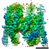
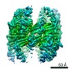
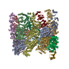
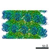
Lidless D386A Mm-cpn in the pre-hydrolysis ATP-bound state
Map data
This is the density map of lidless D386A Mm-cpn in the pre-hydrolysis ATP-bound state.
Sample
Sample: Lidless D386A Mm-cpn variant
Protein or peptide: Methanococcus maripaludis chaperonin
Keywords
Mm-cpn / Chaperonin / ATP-bound
Function / homology
Function and homology information
chaperonin-containing T-complex / ATP-dependent protein folding chaperone / unfolded protein binding / ATP hydrolysis activity / ATP binding / identical protein binding / metal ion binding Similarity search - Function
Journal: Structure / Year: 2011 Title: Cryo-EM structure of a group II chaperonin in the prehydrolysis ATP-bound state leading to lid closure. Authors: Junjie Zhang / Boxue Ma / Frank DiMaio / Nicholai R Douglas / Lukasz A Joachimiak / David Baker / Judith Frydman / Michael Levitt / Wah Chiu / Abstract: Chaperonins are large ATP-driven molecular machines that mediate cellular protein folding. Group II chaperonins use their "built-in lid" to close their central folding chamber. Here we report the ...Chaperonins are large ATP-driven molecular machines that mediate cellular protein folding. Group II chaperonins use their "built-in lid" to close their central folding chamber. Here we report the structure of an archaeal group II chaperonin in its prehydrolysis ATP-bound state at subnanometer resolution using single particle cryo-electron microscopy (cryo-EM). Structural comparison of Mm-cpn in ATP-free, ATP-bound, and ATP-hydrolysis states reveals that ATP binding alone causes the chaperonin to close slightly with a ∼45° counterclockwise rotation of the apical domain. The subsequent ATP hydrolysis drives each subunit to rock toward the folding chamber and to close the lid completely. These motions are attributable to the local interactions of specific active site residues with the nucleotide, the tight couplings between the apical and intermediate domains within the subunit, and the aligned interactions between two subunits across the rings. This mechanism of structural changes in response to ATP is entirely different from those found in group I chaperonins.
History
Deposition
Feb 10, 2011
-
Header (metadata) release
May 11, 2011
-
Map release
May 11, 2011
-
Update
May 7, 2014
-
Current status
May 7, 2014
Processing site: RCSB / Status: Released
-
Structure visualization
Movie
Surface view with section colored by density value
In the structure databanks used in Yorodumi, some data are registered as the other names, "COVID-19 virus" and "2019-nCoV". Here are the details of the virus and the list of structure data.
Jan 31, 2019. EMDB accession codes are about to change! (news from PDBe EMDB page)
EMDB accession codes are about to change! (news from PDBe EMDB page)
The allocation of 4 digits for EMDB accession codes will soon come to an end. Whilst these codes will remain in use, new EMDB accession codes will include an additional digit and will expand incrementally as the available range of codes is exhausted. The current 4-digit format prefixed with “EMD-” (i.e. EMD-XXXX) will advance to a 5-digit format (i.e. EMD-XXXXX), and so on. It is currently estimated that the 4-digit codes will be depleted around Spring 2019, at which point the 5-digit format will come into force.
The EM Navigator/Yorodumi systems omit the EMD- prefix.
Related info.:Q: What is EMD? / ID/Accession-code notation in Yorodumi/EM Navigator
Yorodumi is a browser for structure data from EMDB, PDB, SASBDB, etc.
This page is also the successor to EM Navigator detail page, and also detail information page/front-end page for Omokage search.
The word "yorodu" (or yorozu) is an old Japanese word meaning "ten thousand". "mi" (miru) is to see.
Related info.:EMDB / PDB / SASBDB / Comparison of 3 databanks / Yorodumi Search / Aug 31, 2016. New EM Navigator & Yorodumi / Yorodumi Papers / Jmol/JSmol / Function and homology information / Changes in new EM Navigator and Yorodumi
 Movie
Movie Controller


 Open data
 Basic information
 Map data
 Sample
 Keywords
 Function and homology information
 Methanococcus maripaludis (archaea)
 Authors
 Citation
 Structure visualization
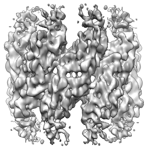
 Downloads & links
 emd_5258_1.png
 http://ftp.pdbj.org/pub/emdb/structures/EMD-5258

 Sample components
 Processing
 Electron microscopy
 FIELD EMISSION GUN