 Movie
Movie Controller


+ Open data

- Basic information
| Entry | Database: EMDB / ID: EMD-5244 | |||||||||
|---|---|---|---|---|---|---|---|---|---|---|
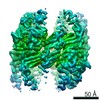
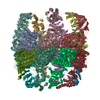
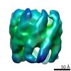
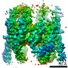
| Title | Mm-cpn D386A with ATP | |||||||||
 Map data Map data | This is the density map of the Mm-cpn D386A mutant with ATP bound. | |||||||||
 Sample Sample |
| |||||||||
 Keywords Keywords | Mm-cpn / maripaludis / chaperonin | |||||||||
| Function / homology |  Function and homology information Function and homology informationchaperonin-containing T-complex / ATP-dependent protein folding chaperone / unfolded protein binding / ATP hydrolysis activity / ATP binding / identical protein binding / metal ion binding Similarity search - Function | |||||||||
| Biological species |  Methanococcus maripaludis (archaea) Methanococcus maripaludis (archaea) | |||||||||
| Method | single particle reconstruction / cryo EM / Resolution: 11.0 Å | |||||||||
 Authors Authors | Douglas NR / Reissmann S / Zhang J / Chen B / Jakana J / Kumar R / Chiu W / Frydman J | |||||||||
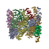
 Citation Citation | Journal: Cell / Year: 2011 Title: Dual action of ATP hydrolysis couples lid closure to substrate release into the group II chaperonin chamber. Authors: Nicholai R Douglas / Stefanie Reissmann / Junjie Zhang / Bo Chen / Joanita Jakana / Ramya Kumar / Wah Chiu / Judith Frydman /  Abstract: Group II chaperonins are ATP-dependent ring-shaped complexes that bind nonnative polypeptides and facilitate protein folding in archaea and eukaryotes. A built-in lid encapsulates substrate proteins ...Group II chaperonins are ATP-dependent ring-shaped complexes that bind nonnative polypeptides and facilitate protein folding in archaea and eukaryotes. A built-in lid encapsulates substrate proteins within the central chaperonin chamber. Here, we describe the fate of the substrate during the nucleotide cycle of group II chaperonins. The chaperonin substrate-binding sites are exposed, and the lid is open in both the ATP-free and ATP-bound prehydrolysis states. ATP hydrolysis has a dual function in the folding cycle, triggering both lid closure and substrate release into the central chamber. Notably, substrate release can occur in the absence of a lid, and lid closure can occur without substrate release. However, productive folding requires both events, so that the polypeptide is released into the confined space of the closed chamber where it folds. Our results show that ATP hydrolysis coordinates the structural and functional determinants that trigger productive folding. | |||||||||
| History |
|
- Structure visualization
| Movie |
Movie viewer |
|---|---|
| Structure viewer | EM map: SurfViewMolmilJmol/JSmol |
| Supplemental images |

- Downloads & links
-EMDB archive
| Map data | emd_5244.map.gz | 23 MB | EMDB map data format | |
|---|---|---|---|---|
| Header (meta data) | emd-5244-v30.xmlemd-5244.xml | 8.2 KB 8.2 KB | Display Display | EMDB header |
| Images | 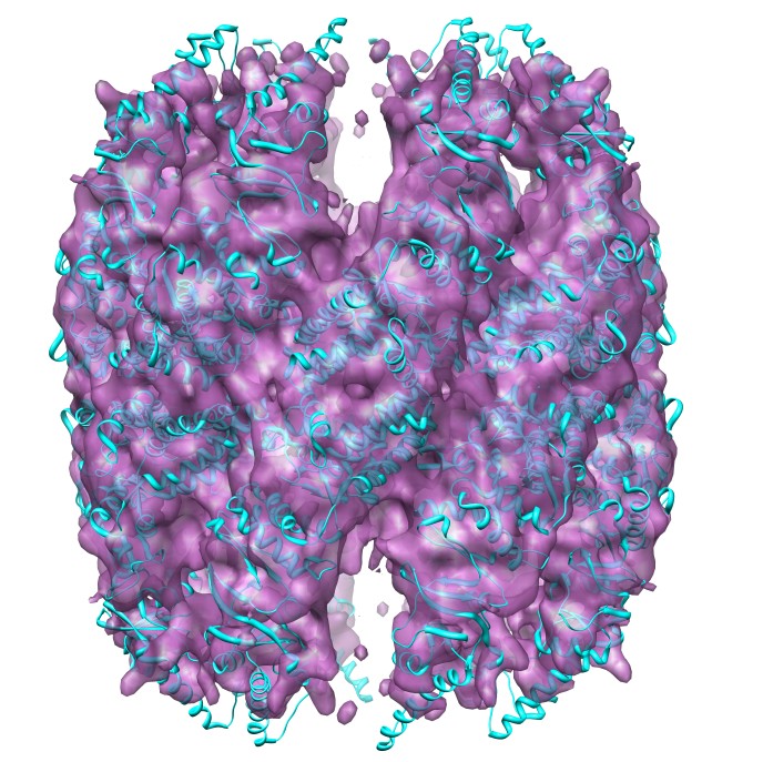 emd_5244_1.jpg emd_5244_1.jpg | 142.1 KB | ||
| Archive directory |  http://ftp.pdbj.org/pub/emdb/structures/EMD-5244ftp://ftp.pdbj.org/pub/emdb/structures/EMD-5244 http://ftp.pdbj.org/pub/emdb/structures/EMD-5244ftp://ftp.pdbj.org/pub/emdb/structures/EMD-5244 | HTTPS FTP |
-Validation report
| Summary document | emd_5244_validation.pdf.gz | 328.5 KB | Display | EMDB validaton report |
|---|---|---|---|---|
| Full document | emd_5244_full_validation.pdf.gz | 328 KB | Display | |
| Data in XML | emd_5244_validation.xml.gz | 6 KB | Display | |
| Arichive directory | https://ftp.pdbj.org/pub/emdb/validation_reports/EMD-5244ftp://ftp.pdbj.org/pub/emdb/validation_reports/EMD-5244 | HTTPS FTP |
-Related structure data
| Related structure data |  3izhMC  5245C  5246C 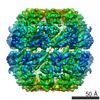 5247C 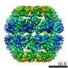 5248C 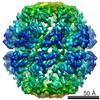 5249C 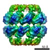 5250C 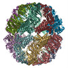 3iziC 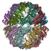 3izjC 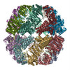 3izkC 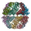 3izlC 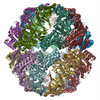 3izmC 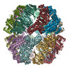 3iznC M: atomic model generated by this map C: citing same article ( |
|---|---|
| Similar structure data |

-Links
| EMDB pages | EMDB (EBI/PDBe) / EMDataResource |
|---|---|
| Related items in Molecule of the Month |

-Map
| File | Download / File: emd_5244.map.gz / Format: CCP4 / Size: 26.4 MB / Type: IMAGE STORED AS FLOATING POINT NUMBER (4 BYTES) | ||||||||||||||||||||||||||||||||||||||||||||||||||||||||||||||||||||
|---|---|---|---|---|---|---|---|---|---|---|---|---|---|---|---|---|---|---|---|---|---|---|---|---|---|---|---|---|---|---|---|---|---|---|---|---|---|---|---|---|---|---|---|---|---|---|---|---|---|---|---|---|---|---|---|---|---|---|---|---|---|---|---|---|---|---|---|---|---|
| Annotation | This is the density map of the Mm-cpn D386A mutant with ATP bound. | ||||||||||||||||||||||||||||||||||||||||||||||||||||||||||||||||||||
| Voxel size | X=Y=Z: 1.81 Å | ||||||||||||||||||||||||||||||||||||||||||||||||||||||||||||||||||||
| Density |
| ||||||||||||||||||||||||||||||||||||||||||||||||||||||||||||||||||||
| Symmetry | Space group: 1 | ||||||||||||||||||||||||||||||||||||||||||||||||||||||||||||||||||||
| Details | EMDB XML:
CCP4 map header:
| ||||||||||||||||||||||||||||||||||||||||||||||||||||||||||||||||||||
-Supplemental data
- Sample components
-Entire : Mm-cpn D386A with ATP
| Entire | Name: Mm-cpn D386A with ATP |
|---|---|
| Components |
|
-Supramolecule #1000: Mm-cpn D386A with ATP
| Supramolecule | Name: Mm-cpn D386A with ATP / type: sample / ID: 1000 / Oligomeric state: 16-mer / Number unique components: 1 |
|---|---|
| Molecular weight | Experimental: 900 KDa / Theoretical: 900 KDa |
-Macromolecule #1: chaperonin
| Macromolecule | Name: chaperonin / type: protein_or_peptide / ID: 1 / Name.synonym: Mm-cpn / Recombinant expression: Yes |
|---|---|
| Source (natural) | Organism: Methanococcus maripaludis (archaea) |
| Molecular weight | Experimental: 900 KDa / Theoretical: 900 KDa |
| Recombinant expression | Organism:  |
-Experimental details
-Structure determination
| Method | cryo EM |
|---|---|
 Processing Processing | single particle reconstruction |
| Aggregation state | particle |
-Sample preparation
| Vitrification | Cryogen name: ETHANE / Chamber humidity: 100 % / Instrument: OTHER / Details: Vitrification instrument: vitrobot |
|---|
- Electron microscopy
| Microscope | JEOL 2010F |
|---|---|
| Image recording | Category: FILM / Film or detector model: GENERIC GATAN (4k x 4k) / Digitization - Scanner: OTHER |
| Electron beam | Acceleration voltage: 200 kV / Electron source:  FIELD EMISSION GUN FIELD EMISSION GUN |
| Electron optics | Illumination mode: FLOOD BEAM / Imaging mode: BRIGHT FIELD |
| Sample stage | Specimen holder: Gatan side entry / Specimen holder model: GATAN LIQUID NITROGEN |
-Image processing
| Final reconstruction | Resolution.type: BY AUTHOR / Resolution: 11.0 Å / Resolution method: FSC 0.5 CUT-OFF / Software - Name: EMAN |
|---|
