 Movie
Movie Controller


+ Open data

- Basic information
| Entry | Database: EMDB / ID: EMD-5224 | |||||||||
|---|---|---|---|---|---|---|---|---|---|---|
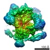
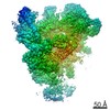
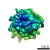
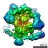
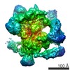
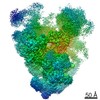
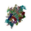
| Title | Human 80S ribosome in situ, untreated Eukaryotic ribosome Eukaryotic ribosome | |||||||||
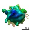
 Map data Map data | This is a map of a tomographic average of a human 80S ribosome in situ | |||||||||
 Sample Sample |
| |||||||||
 Keywords Keywords | human 80S ribosome / puromycin / in situ / cytosol / polysome / polyribosome / protein synthesis / translation / 3D cryoEM / tomography / cellular tomography | |||||||||
| Biological species |  Homo sapiens (human) Homo sapiens (human) | |||||||||
| Method | subtomogram averaging / cryo EM / negative staining / Resolution: 39.0 Å | |||||||||
 Authors Authors | Brandt F / Carlson L-A / Hartl FU / Baumeister W / Grunewald K | |||||||||
 Citation Citation | Journal: Mol Cell / Year: 2010 Title: The three-dimensional organization of polyribosomes in intact human cells. Authors: Florian Brandt / Lars-Anders Carlson / F Ulrich Hartl / Wolfgang Baumeister / Kay Grünewald /  Abstract: Structural studies have provided detailed insights into different functional states of the ribosome and its interaction with factors involved in nascent peptide folding, processing, and targeting. ...Structural studies have provided detailed insights into different functional states of the ribosome and its interaction with factors involved in nascent peptide folding, processing, and targeting. However, how the translational machinery is organized spatially in native cellular environments is not yet well understood. Here we have mapped individual ribosomes in electron tomograms of intact human cells by template matching and determined the average structure of the ribosome in situ. Characteristic features of active ribosomes in the cellular environment were assigned to the tRNA channel, elongation factors, and additional densities near the peptide tunnel. Importantly, the relative spatial configuration of neighboring ribosomes in the cell is clearly nonrandom. The preferred configurations are specific for active polysomes and were largely abrogated in puromycin-treated control cells. The distinct neighbor orientations found in situ resemble configurations of bacterial polysomes in vitro, indicating a conserved supramolecular organization with implications for nascent polypeptide folding. | |||||||||
| History |
|
- Structure visualization
| Movie |
 Movie viewer Movie viewer |
|---|---|
| Structure viewer | EM map: SurfViewMolmilJmol/JSmol |
| Supplemental images |

- Downloads & links
-EMDB archive
| Map data | emd_5224.map.gz | 397.8 KB | EMDB map data format | |
|---|---|---|---|---|
| Header (meta data) | emd-5224-v30.xmlemd-5224.xml | 10.2 KB 10.2 KB | Display Display | EMDB header |
| Images | 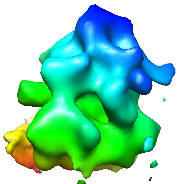 emd_5224_1.jpg emd_5224_1.jpg | 58.6 KB | ||
| Archive directory |  http://ftp.pdbj.org/pub/emdb/structures/EMD-5224ftp://ftp.pdbj.org/pub/emdb/structures/EMD-5224 http://ftp.pdbj.org/pub/emdb/structures/EMD-5224ftp://ftp.pdbj.org/pub/emdb/structures/EMD-5224 | HTTPS FTP |
-Related structure data
-Links
| EMDB pages | EMDB (EBI/PDBe) / EMDataResource |
|---|---|
| Related items in Molecule of the Month |

-Map
| File | Download / File: emd_5224.map.gz / Format: CCP4 / Size: 422.9 KB / Type: IMAGE STORED AS FLOATING POINT NUMBER (4 BYTES) | ||||||||||||||||||||||||||||||||||||||||||||||||||||||||||||||||||||
|---|---|---|---|---|---|---|---|---|---|---|---|---|---|---|---|---|---|---|---|---|---|---|---|---|---|---|---|---|---|---|---|---|---|---|---|---|---|---|---|---|---|---|---|---|---|---|---|---|---|---|---|---|---|---|---|---|---|---|---|---|---|---|---|---|---|---|---|---|---|
| Annotation | This is a map of a tomographic average of a human 80S ribosome in situ | ||||||||||||||||||||||||||||||||||||||||||||||||||||||||||||||||||||
| Voxel size | X=Y=Z: 8.21 Å | ||||||||||||||||||||||||||||||||||||||||||||||||||||||||||||||||||||
| Density |
| ||||||||||||||||||||||||||||||||||||||||||||||||||||||||||||||||||||
| Symmetry | Space group: 1 | ||||||||||||||||||||||||||||||||||||||||||||||||||||||||||||||||||||
| Details | EMDB XML:
CCP4 map header:
| ||||||||||||||||||||||||||||||||||||||||||||||||||||||||||||||||||||
-Supplemental data
- Sample components
-Entire : Human 80S ribosome in situ, untreated
| Entire | Name: Human 80S ribosome in situ, untreatedEukaryotic ribosome |
|---|---|
| Components |
|
-Supramolecule #1000: Human 80S ribosome in situ, untreated
| Supramolecule | Name: Human 80S ribosome in situ, untreated / type: sample / ID: 1000 / Details: Cytosolic ribosomes in situ Oligomeric state: One 80S ribosome within mixed cellular polysomes Number unique components: 1 |
|---|---|
| Molecular weight | Method: Sedimentation, 80S |
-Supramolecule #1: cytosolic 80S ribosome
| Supramolecule | Name: cytosolic 80S ribosome / type: complex / ID: 1 / Recombinant expression: No / Database: NCBI / Ribosome-details: ribosome-eukaryote: ALL |
|---|---|
| Source (natural) | Organism: Homo sapiens (human) / synonym: human |
-Experimental details
-Structure determination
| Method | negative staining, cryo EM |
|---|---|
 Processing Processing | subtomogram averaging |
| Aggregation state | particle |
-Sample preparation
| Buffer | pH: 7.5 Details: Cellular medium, DMEM (Gibco) supplemented with 10% foetal calf serum, 37C and 5% CO2 |
|---|---|
| Staining | Type: NEGATIVE / Details: Cells grown on grids, vitrification |
| Grid | Details: C-flat 2/1, holey carbon gold grid |
| Vitrification | Cryogen name: ETHANE / Chamber temperature: 77 K / Instrument: HOMEMADE PLUNGER Details: Vitrification instrument: plunger. Vitrification carried out in air Method: Blot for 2 s before plunging |
- Electron microscopy
| Microscope | FEI/PHILIPS CM300FEG/T |
|---|---|
| Electron beam | Acceleration voltage: 300 kV / Electron source: FIELD EMISSION GUN |
| Electron optics | Calibrated magnification: 17500 / Illumination mode: FLOOD BEAM / Imaging mode: BRIGHT FIELDBright-field microscopy / Cs: 2 mm / Nominal defocus max: 7.0 µm / Nominal defocus min: 5.0 µm / Nominal magnification: 17500 |
| Specialist optics | Energy filter - Name: GIF2002 / Energy filter - Lower energy threshold: 0.0 eV / Energy filter - Upper energy threshold: 20.0 eV |
| Sample stage | Specimen holder: Side entry liquid nitrogen-cooled cryo specimen holder. Specimen holder model: GATAN LIQUID NITROGEN / Tilt series - Axis1 - Min angle: -65 ° / Tilt series - Axis1 - Max angle: 65 ° |
| Temperature | Min: 77 K / Average: 77 K |
| Alignment procedure | Legacy - Astigmatism: objective lens astigmatism was corrected at 50,000 times magnification |
| Date | Jul 7, 2008 |
| Image recording | Category: CCD / Film or detector model: GATAN MULTISCAN / Average electron dose: 80 e/Å2 |
-Image processing
| Final 3D classification | Number classes: 1 |
|---|---|
| Final reconstruction | Algorithm: OTHER / Resolution.type: BY AUTHOR / Resolution: 39.0 Å / Resolution method: FSC 0.5 CUT-OFF / Software - Name: EM / Details: exact weighting / Number subtomograms used: 1911 |
| Details | Individual particle subvolumes were automatically selected by CCC threshold. |
