 Movie
Movie Controller


+ Open data

- Basic information
| Entry | Database: EMDB / ID: EMD-5209 | |||||||||
|---|---|---|---|---|---|---|---|---|---|---|
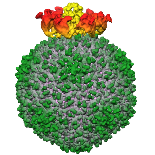
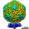
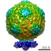
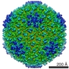
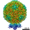
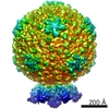
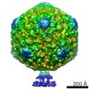
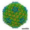
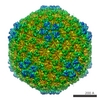
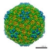
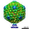
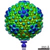
| Title | Epsilon15 asymmetric reconstruction using conventional cryo-EM | |||||||||
 Map data Map data | asymmetric reconstruction of epsilon15 from conventional cryoEM particles | |||||||||
 Sample Sample |
| |||||||||
 Keywords Keywords |  Electron microscopy / phase plate / Bacteriophage / epsilon15 Electron microscopy / phase plate / Bacteriophage / epsilon15 | |||||||||
| Biological species |  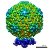 epsilon15 (virus) epsilon15 (virus) | |||||||||
| Method | single particle reconstruction / cryo EM / Resolution: 13.0 Å | |||||||||
 Authors Authors | Murata K / Liu X / Danev R / Jakana J / Schmid MF / King J / Nagayama K / Chiu W | |||||||||
 Citation Citation | Journal: Structure / Year: 2010 Title: Zernike phase contrast cryo-electron microscopy and tomography for structure determination at nanometer and subnanometer resolutions. Authors: Kazuyoshi Murata / Xiangan Liu / Radostin Danev / Joanita Jakana / Michael F Schmid / Jonathan King / Kuniaki Nagayama / Wah Chiu /  Abstract: Zernike phase contrast cryo-electron microscopy (ZPC-cryoEM) is an emerging technique that is capable of producing higher image contrast than conventional cryoEM. By combining this technique with ...Zernike phase contrast cryo-electron microscopy (ZPC-cryoEM) is an emerging technique that is capable of producing higher image contrast than conventional cryoEM. By combining this technique with advanced image processing methods, we achieved subnanometer resolution for two biological specimens: 2D bacteriorhodopsin crystal and epsilon15 bacteriophage. For an asymmetric reconstruction of epsilon15 bacteriophage, ZPC-cryoEM can reduce the required amount of data by a factor of approximately 3, compared with conventional cryoEM. The reconstruction was carried out to 13 A resolution without the need to correct the contrast transfer function. New structural features at the portal vertex of the epsilon15 bacteriophage are revealed in this reconstruction. Using ZPC cryo-electron tomography (ZPC-cryoET), a similar level of data reduction and higher resolution structures of epsilon15 bacteriophage can be obtained relative to conventional cryoET. These results show quantitatively the benefits of ZPC-cryoEM and ZPC-cryoET for structural determinations of macromolecular machines at nanometer and subnanometer resolutions. | |||||||||
| History |
|
- Structure visualization
| Movie |
 Movie viewer Movie viewer |
|---|---|
| Structure viewer | EM map: SurfViewMolmilJmol/JSmol |
| Supplemental images |
- Downloads & links
-EMDB archive
| Map data | emd_5209.map.gz | 97.7 MB | EMDB map data format | |
|---|---|---|---|---|
| Header (meta data) | emd-5209-v30.xmlemd-5209.xml | 9.6 KB 9.6 KB | Display Display | EMDB header |
| Images |  emd=5209_1.png emd=5209_1.png | 317.6 KB | ||
| Archive directory |  http://ftp.pdbj.org/pub/emdb/structures/EMD-5209ftp://ftp.pdbj.org/pub/emdb/structures/EMD-5209 http://ftp.pdbj.org/pub/emdb/structures/EMD-5209ftp://ftp.pdbj.org/pub/emdb/structures/EMD-5209 | HTTPS FTP |
-Related structure data
-Links
| EMDB pages | EMDB (EBI/PDBe) / EMDataResource |
|---|
-Map
| File | Download / File: emd_5209.map.gz / Format: CCP4 / Size: 711.9 MB / Type: IMAGE STORED AS FLOATING POINT NUMBER (4 BYTES) | ||||||||||||||||||||||||||||||||||||||||||||||||||||||||||||||||||||
|---|---|---|---|---|---|---|---|---|---|---|---|---|---|---|---|---|---|---|---|---|---|---|---|---|---|---|---|---|---|---|---|---|---|---|---|---|---|---|---|---|---|---|---|---|---|---|---|---|---|---|---|---|---|---|---|---|---|---|---|---|---|---|---|---|---|---|---|---|---|
| Annotation | asymmetric reconstruction of epsilon15 from conventional cryoEM particles | ||||||||||||||||||||||||||||||||||||||||||||||||||||||||||||||||||||
| Voxel size | X=Y=Z: 1.95 Å | ||||||||||||||||||||||||||||||||||||||||||||||||||||||||||||||||||||
| Density |
| ||||||||||||||||||||||||||||||||||||||||||||||||||||||||||||||||||||
| Symmetry | Space group: 1 | ||||||||||||||||||||||||||||||||||||||||||||||||||||||||||||||||||||
| Details | EMDB XML:
CCP4 map header:
| ||||||||||||||||||||||||||||||||||||||||||||||||||||||||||||||||||||
-Supplemental data
- Sample components
-Entire : Epsilon15
| Entire | Name: Epsilon15 (virus) |
|---|---|
| Components |
|
-Supramolecule #1000: Epsilon15
| Supramolecule | Name: Epsilon15 / type: sample / ID: 1000 / Details: The sample was monodisperse / Number unique components: 6 |
|---|---|
| Molecular weight | Theoretical: 500 KDa |
-Supramolecule #1: epsilon15
| Supramolecule | Name: epsilon15 / type: virus / ID: 1 / Name.synonym: bacteriophage / Sci species name: epsilon15 / Database: NCBI / Virus type: VIRION / Virus isolate: STRAIN / Virus enveloped: No / Virus empty: No / Syn species name: bacteriophage |
|---|---|
| Host (natural) | Organism:  Salmonella (bacteria) / synonym: BACTERIA(EUBACTERIA) Salmonella (bacteria) / synonym: BACTERIA(EUBACTERIA) |
| Virus shell | Shell ID: 1 / Diameter: 700 Å / T number (triangulation number): 7 |
-Experimental details
-Structure determination
| Method | cryo EM |
|---|---|
 Processing Processing | single particle reconstruction |
| Aggregation state | particle |
-Sample preparation
| Buffer | pH: 7.5 / Details: 10 mM Tris pH7.5, 25 mM NaCl and 5 mM MgCl2 |
|---|---|
| Grid | Details: Quantifoil R2/2 grid |
| Vitrification | Cryogen name: ETHANE / Instrument: OTHER / Details: Vitrification instrument: vitrobot / Method: Blot for 2 seconds before plunging |
- Electron microscopy
| Microscope | JEOL 2200FS |
|---|---|
| Electron beam | Acceleration voltage: 200 kV / Electron source: FIELD EMISSION GUN |
| Electron optics | Calibrated magnification: 75000 / Illumination mode: FLOOD BEAM / Imaging mode: BRIGHT FIELDBright-field microscopy / Cs: 4.3 mm / Nominal magnification: 75000 |
| Specialist optics | Energy filter - Name: JEOL / Energy filter - Lower energy threshold: 0.0 eV / Energy filter - Upper energy threshold: 20.0 eV |
| Sample stage | Specimen holder: Eucentric / Specimen holder model: GATAN LIQUID NITROGEN |
| Temperature | Average: 90 K |
| Details | Weak beam illumination |
| Date | Dec 1, 2009 |
| Image recording | Category: CCD / Film or detector model: GENERIC TVIPS (4k x 4k) / Number real images: 722 / Average electron dose: 20 e/Å2 / Bits/pixel: 16 |
-Image processing
| CTF correction | Details: each micrograph |
|---|---|
| Final reconstruction | Algorithm: OTHER / Resolution.type: BY AUTHOR / Resolution: 13.0 Å / Resolution method: FSC 0.5 CUT-OFF / Software - Name: MPSA / Details: the map was reconstructed with ctf correction. / Number images used: 17800 |
| Details | The particles were selected using the consistency criterion of MPSA |
