 Movie
Movie Controller


+ Open data

- Basic information
| Entry | Database: EMDB / ID: EMD-5199 | |||||||||
|---|---|---|---|---|---|---|---|---|---|---|
| Title | Atomic model of an infectious rotavirus particle | |||||||||
 Map data Map data | Infectious rotavirus particle. Center of the virus is at 0,0,0 and the map is a quadrant of the actual map. | |||||||||
 Sample Sample |
| |||||||||
 Keywords Keywords | Rotavirus / Triple Layered Particle / Near Atomic Resolution / VP4 / VP7 / Double layered particle / de novo / Infectious / Virus | |||||||||
| Function / homology |  Function and homology information Function and homology informationviral intermediate capsid / host cell endoplasmic reticulum lumen / T=13 icosahedral viral capsid / T=2 icosahedral viral capsid / host cell rough endoplasmic reticulum / viral inner capsid / host cytoskeleton / viral outer capsid / permeabilization of host organelle membrane involved in viral entry into host cell / symbiont entry into host cell via permeabilization of inner membrane ...viral intermediate capsid / host cell endoplasmic reticulum lumen / T=13 icosahedral viral capsid / T=2 icosahedral viral capsid / host cell rough endoplasmic reticulum / viral inner capsid / host cytoskeleton / viral outer capsid / permeabilization of host organelle membrane involved in viral entry into host cell / symbiont entry into host cell via permeabilization of inner membrane / host cell endoplasmic reticulum-Golgi intermediate compartment / viral nucleocapsid / receptor-mediated virion attachment to host cell / host cell surface receptor binding / fusion of virus membrane with host plasma membrane / viral envelope / virion attachment to host cell / host cell plasma membrane / structural molecule activity / RNA binding / membrane / metal ion binding Similarity search - Function | |||||||||
| Biological species | Rhesus Rotavirus (RRV) | |||||||||
| Method | single particle reconstruction / cryo EM / Resolution: 3.8 Å | |||||||||
 Authors Authors | Settembre EC / Chen JZ / Dormitzer PR / Grigorieff N / Harrison SC | |||||||||
 Citation Citation | Journal: EMBO J / Year: 2011 Title: Atomic model of an infectious rotavirus particle. Authors: Ethan C Settembre / James Z Chen / Philip R Dormitzer / Nikolaus Grigorieff / Stephen C Harrison /  Abstract: Non-enveloped viruses of different types have evolved distinct mechanisms for penetrating a cellular membrane during infection. Rotavirus penetration appears to occur by a process resembling ...Non-enveloped viruses of different types have evolved distinct mechanisms for penetrating a cellular membrane during infection. Rotavirus penetration appears to occur by a process resembling enveloped-virus fusion: membrane distortion linked to conformational changes in a viral protein. Evidence for such a mechanism comes from crystallographic analyses of fragments of VP4, the rotavirus-penetration protein, and infectivity analyses of structure-based VP4 mutants. We describe here the structure of an infectious rotavirus particle determined by electron cryomicroscopy (cryoEM) and single-particle analysis at about 4.3 Å resolution. The cryoEM image reconstruction permits a nearly complete trace of the VP4 polypeptide chain, including the positions of most side chains. It shows how the two subfragments of VP4 (VP8(*) and VP5(*)) retain their association after proteolytic cleavage, reveals multiple structural roles for the β-barrel domain of VP5(*), and specifies interactions of VP4 with other capsid proteins. The virion model allows us to integrate structural and functional information into a coherent mechanism for rotavirus entry. | |||||||||
| History |
|
- Structure visualization
| Movie |
Movie viewer |
|---|---|
| Structure viewer | EM map: SurfViewMolmilJmol/JSmol |
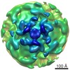
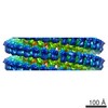
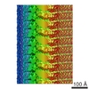
| Supplemental images |

- Downloads & links
-EMDB archive
| Map data | emd_5199.map.gz | 374.1 MB | EMDB map data format | |
|---|---|---|---|---|
| Header (meta data) | emd-5199-v30.xmlemd-5199.xml | 11.2 KB 11.2 KB | Display Display | EMDB header |
| Images | 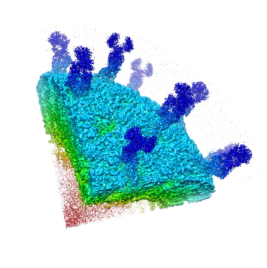 emd_5199_1.jpg emd_5199_1.jpg | 213.2 KB | ||
| Archive directory |  http://ftp.pdbj.org/pub/emdb/structures/EMD-5199ftp://ftp.pdbj.org/pub/emdb/structures/EMD-5199 http://ftp.pdbj.org/pub/emdb/structures/EMD-5199ftp://ftp.pdbj.org/pub/emdb/structures/EMD-5199 | HTTPS FTP |
-Validation report
| Summary document | emd_5199_validation.pdf.gz | 340.2 KB | Display | EMDB validaton report |
|---|---|---|---|---|
| Full document | emd_5199_full_validation.pdf.gz | 339.8 KB | Display | |
| Data in XML | emd_5199_validation.xml.gz | 7.8 KB | Display | |
| Arichive directory | https://ftp.pdbj.org/pub/emdb/validation_reports/EMD-5199ftp://ftp.pdbj.org/pub/emdb/validation_reports/EMD-5199 | HTTPS FTP |
-Related structure data
| Related structure data |  4v7qMC M: atomic model generated by this map C: citing same article ( |
|---|---|
| Similar structure data |

-Links
| EMDB pages | EMDB (EBI/PDBe) / EMDataResource |
|---|---|
| Related items in Molecule of the Month |


-Map
| File | Download / File: emd_5199.map.gz / Format: CCP4 / Size: 465.7 MB / Type: IMAGE STORED AS FLOATING POINT NUMBER (4 BYTES) | ||||||||||||||||||||||||||||||||||||||||||||||||||||||||||||||||||||
|---|---|---|---|---|---|---|---|---|---|---|---|---|---|---|---|---|---|---|---|---|---|---|---|---|---|---|---|---|---|---|---|---|---|---|---|---|---|---|---|---|---|---|---|---|---|---|---|---|---|---|---|---|---|---|---|---|---|---|---|---|---|---|---|---|---|---|---|---|---|
| Annotation | Infectious rotavirus particle. Center of the virus is at 0,0,0 and the map is a quadrant of the actual map. | ||||||||||||||||||||||||||||||||||||||||||||||||||||||||||||||||||||
| Voxel size | X=Y=Z: 1.23 Å | ||||||||||||||||||||||||||||||||||||||||||||||||||||||||||||||||||||
| Density |
| ||||||||||||||||||||||||||||||||||||||||||||||||||||||||||||||||||||
| Symmetry | Space group: 1 | ||||||||||||||||||||||||||||||||||||||||||||||||||||||||||||||||||||
| Details | EMDB XML:
CCP4 map header:
| ||||||||||||||||||||||||||||||||||||||||||||||||||||||||||||||||||||
-Supplemental data
- Sample components
-Entire : Rotavirus Triple Layered Particle
| Entire | Name: Rotavirus Triple Layered Particle |
|---|---|
| Components |
|
-Supramolecule #1000: Rotavirus Triple Layered Particle
| Supramolecule | Name: Rotavirus Triple Layered Particle / type: sample / ID: 1000 / Details: The sample was monodisperse / Oligomeric state: Full icosahedral Infectious Particle / Number unique components: 1 |
|---|---|
| Molecular weight | Experimental: 100 MDa / Theoretical: 100 MDa Method: Calculation of Protein Mass alone, no genome included. |
-Supramolecule #1: Rhesus Rotavirus (RRV)
| Supramolecule | Name: Rhesus Rotavirus (RRV) / type: virus / ID: 1 / Name.synonym: Rotavirus Details: The entire particle with genome was measured (the molecular weight is based on protein alone). Sci species name: Rhesus Rotavirus (RRV) / Database: NCBI / Virus type: VIRION / Virus isolate: STRAIN / Virus enveloped: No / Virus empty: No / Syn species name: Rotavirus |
|---|---|
| Host (natural) | Organism:  Homo sapiens (human) / synonym: VERTEBRATES Homo sapiens (human) / synonym: VERTEBRATES |
| Molecular weight | Experimental: 100 MDa / Theoretical: 100 MDa |
| Virus shell | Shell ID: 1 / Name: VP7 / Diameter: 800 Å / T number (triangulation number): 13 |
-Experimental details
-Structure determination
| Method | cryo EM |
|---|---|
 Processing Processing | single particle reconstruction |
| Aggregation state | particle |
-Sample preparation
| Concentration | 1 mg/mL |
|---|---|
| Buffer | pH: 7.5 / Details: 10 mM Tris-HCL |
| Grid | Details: 200 mesh Carbon |
| Vitrification | Cryogen name: ETHANE / Chamber humidity: 90 % / Chamber temperature: 90 K / Instrument: HOMEMADE PLUNGER Details: Vitrification instrument: manual plunger. in normal coldroom environment Method: front blotting for 3s before plunging |
- Electron microscopy
| Microscope | FEI TECNAI F30 |
|---|---|
| Temperature | Min: 90 K / Max: 90 K / Average: 90 K |
| Details | Cut-plate film holders to reduce electron back-scattering |
| Date | Mar 1, 2008 |
| Image recording | Category: FILM / Film or detector model: KODAK SO-163 FILM / Digitization - Scanner: ZEISS SCAI / Digitization - Sampling interval: 7.0 µm / Number real images: 110 / Average electron dose: 20 e/Å2 / Camera length: 800 / Details: 4,187 particles in the dataset / Od range: 1.2 / Bits/pixel: 8 |
| Electron beam | Acceleration voltage: 300 kV / Electron source:  FIELD EMISSION GUN FIELD EMISSION GUN |
| Electron optics | Calibrated magnification: 56772 / Illumination mode: FLOOD BEAM / Imaging mode: BRIGHT FIELD / Cs: 2.0 mm / Nominal defocus max: 3.0 µm / Nominal defocus min: 1.2 µm / Nominal magnification: 59000 |
| Sample stage | Specimen holder: Eucentric / Specimen holder model: GATAN LIQUID NITROGEN |
| Experimental equipment |  Model: Tecnai F30 / Image courtesy: FEI Company |
