 Movie
Movie Controller


+ Open data

- Basic information
| Entry | Database: EMDB / ID: EMD-5140 | |||||||||
|---|---|---|---|---|---|---|---|---|---|---|
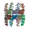
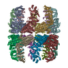
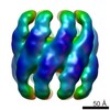
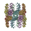
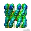
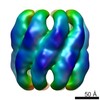
| Title | Lidless Mm-cpn in the open state | |||||||||
 Map data Map data | This is a map of the lidless Mm-cpn in the nucleotide-free open state. | |||||||||
 Sample Sample |
| |||||||||
 Keywords Keywords | Group II chaperonin /  Protein Folding / Mm-cpn / Single Particle Reconstruction / Methanococcus maripaludis / Chaperone Protein Folding / Mm-cpn / Single Particle Reconstruction / Methanococcus maripaludis / Chaperone | |||||||||
| Function / homology |  Function and homology information Function and homology informationATP-dependent protein folding chaperone / unfolded protein binding / ATP hydrolysis activity / ATP binding / identical protein bindingSimilarity search - Function | |||||||||
| Biological species |  Methanococcus maripaludis (archaea) Methanococcus maripaludis (archaea) | |||||||||
| Method | single particle reconstruction / cryo EM / Resolution: 8.0 Å | |||||||||
 Authors Authors | Zhang J / Baker ML / Schroeder G / Douglas NR / Reissmann S / Jakana J / Dougherty M / Fu CJ / Levitt M / Ludtke SJ ...Zhang J / Baker ML / Schroeder G / Douglas NR / Reissmann S / Jakana J / Dougherty M / Fu CJ / Levitt M / Ludtke SJ / Frydman J / Chiu W | |||||||||
 Citation Citation | Journal: Nature / Year: 2010 Title: Mechanism of folding chamber closure in a group II chaperonin. Authors: Junjie Zhang / Matthew L Baker / Gunnar F Schröder / Nicholai R Douglas / Stefanie Reissmann / Joanita Jakana / Matthew Dougherty / Caroline J Fu / Michael Levitt / Steven J Ludtke / Judith ...Authors: Junjie Zhang / Matthew L Baker / Gunnar F Schröder / Nicholai R Douglas / Stefanie Reissmann / Joanita Jakana / Matthew Dougherty / Caroline J Fu / Michael Levitt / Steven J Ludtke / Judith Frydman / Wah Chiu /  Abstract: Group II chaperonins are essential mediators of cellular protein folding in eukaryotes and archaea. These oligomeric protein machines, approximately 1 megadalton, consist of two back-to-back rings ...Group II chaperonins are essential mediators of cellular protein folding in eukaryotes and archaea. These oligomeric protein machines, approximately 1 megadalton, consist of two back-to-back rings encompassing a central cavity that accommodates polypeptide substrates. Chaperonin-mediated protein folding is critically dependent on the closure of a built-in lid, which is triggered by ATP hydrolysis. The structural rearrangements and molecular events leading to lid closure are still unknown. Here we report four single particle cryo-electron microscopy (cryo-EM) structures of Mm-cpn, an archaeal group II chaperonin, in the nucleotide-free (open) and nucleotide-induced (closed) states. The 4.3 A resolution of the closed conformation allowed building of the first ever atomic model directly from the single particle cryo-EM density map, in which we were able to visualize the nucleotide and more than 70% of the side chains. The model of the open conformation was obtained by using the deformable elastic network modelling with the 8 A resolution open-state cryo-EM density restraints. Together, the open and closed structures show how local conformational changes triggered by ATP hydrolysis lead to an alteration of intersubunit contacts within and across the rings, ultimately causing a rocking motion that closes the ring. Our analyses show that there is an intricate and unforeseen set of interactions controlling allosteric communication and inter-ring signalling, driving the conformational cycle of group II chaperonins. Beyond this, we anticipate that our methodology of combining single particle cryo-EM and computational modelling will become a powerful tool in the determination of atomic details involved in the dynamic processes of macromolecular machines in solution. | |||||||||
| History |
|
- Structure visualization
| Movie |
Movie viewer |
|---|---|
| Structure viewer | EM map: SurfViewMolmilJmol/JSmol |
| Supplemental images |

- Downloads & links
-EMDB archive
| Map data | emd_5140.map.gz | 10.4 MB | EMDB map data format | |
|---|---|---|---|---|
| Header (meta data) | emd-5140-v30.xmlemd-5140.xml | 10.1 KB 10.1 KB | Display Display | EMDB header |
| Images | 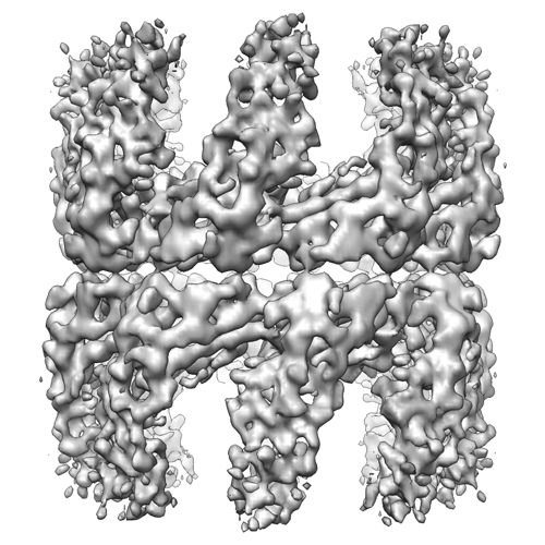 emd_5140_1.png emd_5140_1.png | 219.6 KB | ||
| Archive directory |  http://ftp.pdbj.org/pub/emdb/structures/EMD-5140ftp://ftp.pdbj.org/pub/emdb/structures/EMD-5140 http://ftp.pdbj.org/pub/emdb/structures/EMD-5140ftp://ftp.pdbj.org/pub/emdb/structures/EMD-5140 | HTTPS FTP |
-Related structure data
| Related structure data | 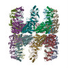 3iyfMC 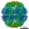 5137C 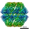 5138C 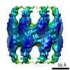 5139C 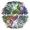 3losC M: atomic model generated by this map C: citing same article ( |
|---|---|
| Similar structure data |

-Links
| EMDB pages | EMDB (EBI/PDBe) / EMDataResource |
|---|---|
| Related items in Molecule of the Month |

-Map
| File | Download / File: emd_5140.map.gz / Format: CCP4 / Size: 51.5 MB / Type: IMAGE STORED AS FLOATING POINT NUMBER (4 BYTES) | ||||||||||||||||||||||||||||||||||||||||||||||||||||||||||||||||||||
|---|---|---|---|---|---|---|---|---|---|---|---|---|---|---|---|---|---|---|---|---|---|---|---|---|---|---|---|---|---|---|---|---|---|---|---|---|---|---|---|---|---|---|---|---|---|---|---|---|---|---|---|---|---|---|---|---|---|---|---|---|---|---|---|---|---|---|---|---|---|
| Annotation | This is a map of the lidless Mm-cpn in the nucleotide-free open state. | ||||||||||||||||||||||||||||||||||||||||||||||||||||||||||||||||||||
| Voxel size | X=Y=Z: 1.33 Å | ||||||||||||||||||||||||||||||||||||||||||||||||||||||||||||||||||||
| Density |
| ||||||||||||||||||||||||||||||||||||||||||||||||||||||||||||||||||||
| Symmetry | Space group: 1 | ||||||||||||||||||||||||||||||||||||||||||||||||||||||||||||||||||||
| Details | EMDB XML:
CCP4 map header:
| ||||||||||||||||||||||||||||||||||||||||||||||||||||||||||||||||||||
-Supplemental data
- Sample components
-Entire : Lidless Mm-cpn
| Entire | Name: Lidless Mm-cpn |
|---|---|
| Components |
|
-Supramolecule #1000: Lidless Mm-cpn
| Supramolecule | Name: Lidless Mm-cpn / type: sample / ID: 1000 / Oligomeric state: 16-mer / Number unique components: 1 |
|---|---|
| Molecular weight | Experimental: 960 KDa / Theoretical: 960 KDa |
-Macromolecule #1: Lidless Methanococcus maripaludis chaperonin
| Macromolecule | Name: Lidless Methanococcus maripaludis chaperonin / type: protein_or_peptide / ID: 1 / Name.synonym: Lidless Mm-cpn / Number of copies: 16 / Oligomeric state: 16-mer / Recombinant expression: Yes |
|---|---|
| Source (natural) | Organism: Methanococcus maripaludis (archaea) |
| Molecular weight | Experimental: 960 KDa / Theoretical: 960 KDa |
| Recombinant expression | Organism:  Escherichia coli (E. coli) Escherichia coli (E. coli) |
-Experimental details
-Structure determination
| Method | cryo EM |
|---|---|
 Processing Processing | single particle reconstruction |
| Aggregation state | particle |
-Sample preparation
| Vitrification | Cryogen name: ETHANE / Chamber humidity: 100 % / Instrument: OTHER / Details: Vitrification instrument: Vitrobot / Method: 1 blot 3 seconds |
|---|
- Electron microscopy
| Microscope | JEOL 3200FSC |
|---|---|
| Electron beam | Acceleration voltage: 300 kV / Electron source: FIELD EMISSION GUN |
| Electron optics | Calibrated magnification: 112000 / Illumination mode: FLOOD BEAM / Imaging mode: BRIGHT FIELDBright-field microscopy / Cs: 4.1 mm / Nominal defocus max: 3.0 µm / Nominal defocus min: 1.0 µm / Nominal magnification: 80000 |
| Specialist optics | Energy filter - Name: in column omega filter / Energy filter - Lower energy threshold: 0.0 eV / Energy filter - Upper energy threshold: 10.0 eV |
| Sample stage | Specimen holder: side-entry / Specimen holder model: JEOL 3200FSC CRYOHOLDER |
| Temperature | Average: 100 K |
| Alignment procedure | Legacy - Astigmatism: objective lens astigmatism was corrected at 100,000 times magnification |
| Image recording | Category: CCD / Film or detector model: GENERIC GATAN (4k x 4k) / Average electron dose: 20 e/Å2 |
-Image processing
| CTF correction | Details: Each micrograph |
|---|---|
| Final reconstruction | Algorithm: OTHER / Resolution.type: BY AUTHOR / Resolution: 8.0 Å / Resolution method: FSC 0.5 CUT-OFF / Software - Name: EMAN |
