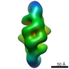
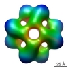
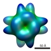
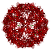
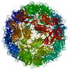
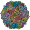
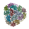
ジャーナル: Proc Natl Acad Sci U S A / 年: 2009 タイトル: Survey of large protein complexes in D. vulgaris reveals great structural diversity. 著者: Bong-Gyoon Han / Ming Dong / Haichuan Liu / Lauren Camp / Jil Geller / Mary Singer / Terry C Hazen / Megan Choi / H Ewa Witkowska / David A Ball / Dieter Typke / Kenneth H Downing / Maxim ...著者: Bong-Gyoon Han / Ming Dong / Haichuan Liu / Lauren Camp / Jil Geller / Mary Singer / Terry C Hazen / Megan Choi / H Ewa Witkowska / David A Ball / Dieter Typke / Kenneth H Downing / Maxim Shatsky / Steven E Brenner / John-Marc Chandonia / Mark D Biggin / Robert M Glaeser / 要旨: An unbiased survey has been made of the stable, most abundant multi-protein complexes in Desulfovibrio vulgaris Hildenborough (DvH) that are larger than Mr approximately 400 k. The quaternary ...An unbiased survey has been made of the stable, most abundant multi-protein complexes in Desulfovibrio vulgaris Hildenborough (DvH) that are larger than Mr approximately 400 k. The quaternary structures for 8 of the 16 complexes purified during this work were determined by single-particle reconstruction of negatively stained specimens, a success rate approximately 10 times greater than that of previous "proteomic" screens. In addition, the subunit compositions and stoichiometries of the remaining complexes were determined by biochemical methods. Our data show that the structures of only two of these large complexes, out of the 13 in this set that have recognizable functions, can be modeled with confidence based on the structures of known homologs. These results indicate that there is significantly greater variability in the way that homologous prokaryotic macromolecular complexes are assembled than has generally been appreciated. As a consequence, we suggest that relying solely on previously determined quaternary structures for homologous proteins may not be sufficient to properly understand their role in another cell of interest.
生物種: Desulfovibrio vulgaris str. Hildenborough (バクテリア) 細胞中の位置: cytoplasmic
分子量
実験値: 996 KDa / 理論値: 996 KDa
-
実験情報
-
構造解析
手法
ネガティブ染色法
解析
単粒子再構成法
試料の集合状態
particle
-
試料調製
濃度
0.3 mg/mL
緩衝液
pH: 7 / 詳細: 10 mM HEPES buffer
染色
タイプ: NEGATIVE 詳細: Three microliter of 2% w/v uranyl acetate stain was applied to the EM grid for 1 min.
グリッド
詳細: carbon-coated and glow-discharged 300 mesh copper grid
凍結
凍結剤: NONE / 装置: OTHER
-
電子顕微鏡法
顕微鏡
JEOL 4000EX
温度
平均: 293 K
アライメント法
Legacy - 非点収差: objective lens astigmatism was corrected at 60,000 times magnification
日付
2008年7月8日
撮影
カテゴリ: FILM / フィルム・検出器のモデル: KODAK SO-163 FILM デジタル化 - スキャナー: NIKON SUPER COOLSCAN 9000 デジタル化 - サンプリング間隔: 6.35 µm / 実像数: 23 / 平均電子線量: 17 e/Å2 詳細: The images were scanned with a resolution of 6.35 micro m per pixel and later averaged 2 fold in each direction. ビット/ピクセル: 8
 ムービー
ムービー コントローラー


 データを開く
 基本情報
基本情報 マップデータ
 試料
試料 キーワード
キーワード Desulfovibrio vulgaris str. Hildenborough (バクテリア)
 データ登録者
 引用
引用
 構造の表示
構造の表示 ムービービューア
 ダウンロードとリンク
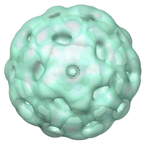 emd_5042_1.jpg
 http://ftp.pdbj.org/pub/emdb/structures/EMD-5042


 試料の構成要素
 解析
解析 電子顕微鏡法
 FIELD EMISSION GUN