 Movie
Movie Controller


+ Open data

- Basic information
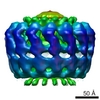
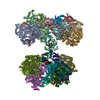
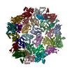
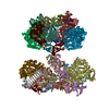
| Entry | Database: EMDB / ID: EMD-5034 | |||||||||
|---|---|---|---|---|---|---|---|---|---|---|
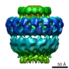
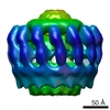
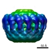
| Title | Structure of a type IV secretion system core complex | |||||||||
 Map data Map data | volume | |||||||||
 Sample Sample |
| |||||||||
 Keywords Keywords | bacterial secretion / type IV secretion / vir / tra | |||||||||
| Method | single particle reconstruction / cryo EM / Resolution: 20.0 Å | |||||||||
 Authors Authors | Fronzes R / Schafer E / Wang L / Saibil H / Orlova E / Waksman G | |||||||||
 Citation Citation | Journal: Science / Year: 2009 Title: Structure of a type IV secretion system core complex. Authors: Rémi Fronzes / Eva Schäfer / Luchun Wang / Helen R Saibil / Elena V Orlova / Gabriel Waksman /  Abstract: Type IV secretion systems (T4SSs) are important virulence factors used by Gram-negative bacterial pathogens to inject effectors into host cells or to spread plasmids harboring antibiotic resistance ...Type IV secretion systems (T4SSs) are important virulence factors used by Gram-negative bacterial pathogens to inject effectors into host cells or to spread plasmids harboring antibiotic resistance genes. We report the 15 angstrom resolution cryo-electron microscopy structure of the core complex of a T4SS. The core complex is composed of three proteins, each present in 14 copies and forming a approximately 1.1-megadalton two-chambered, double membrane-spanning channel. The structure is double-walled, with each component apparently spanning a large part of the channel. The complex is open on the cytoplasmic side and constricted on the extracellular side. Overall, the T4SS core complex structure is different in both architecture and composition from the other known double membrane-spanning secretion system that has been structurally characterized. | |||||||||
| History |
|
- Structure visualization
| Movie |
 Movie viewer Movie viewer |
|---|---|
| Structure viewer | EM map: SurfViewMolmilJmol/JSmol |
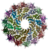
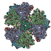
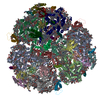
| Supplemental images |

- Downloads & links
-EMDB archive
| Map data | emd_5034.map.gz | 1.1 MB | EMDB map data format | |
|---|---|---|---|---|
| Header (meta data) | emd-5034-v30.xmlemd-5034.xml | 11.9 KB 11.9 KB | Display Display | EMDB header |
| Images | 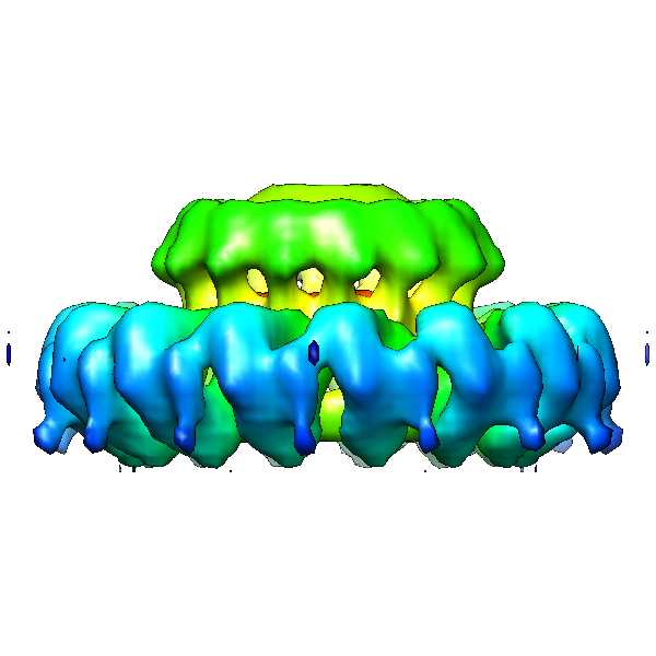 emd_5034_1.png emd_5034_1.png | 126.6 KB | ||
| Archive directory |  http://ftp.pdbj.org/pub/emdb/structures/EMD-5034ftp://ftp.pdbj.org/pub/emdb/structures/EMD-5034 http://ftp.pdbj.org/pub/emdb/structures/EMD-5034ftp://ftp.pdbj.org/pub/emdb/structures/EMD-5034 | HTTPS FTP |
-Validation report
| Summary document | emd_5034_validation.pdf.gz | 78 KB | Display | EMDB validaton report |
|---|---|---|---|---|
| Full document | emd_5034_full_validation.pdf.gz | 77.1 KB | Display | |
| Data in XML | emd_5034_validation.xml.gz | 493 B | Display | |
| Arichive directory | https://ftp.pdbj.org/pub/emdb/validation_reports/EMD-5034ftp://ftp.pdbj.org/pub/emdb/validation_reports/EMD-5034 | HTTPS FTP |
-Related structure data
-Links
| EMDB pages | EMDB (EBI/PDBe) / EMDataResource |
|---|
-Map
| File | Download / File: emd_5034.map.gz / Format: CCP4 / Size: 15.3 MB / Type: IMAGE STORED AS FLOATING POINT NUMBER (4 BYTES) | ||||||||||||||||||||||||||||||||||||||||||||||||||||||||||||||||||||
|---|---|---|---|---|---|---|---|---|---|---|---|---|---|---|---|---|---|---|---|---|---|---|---|---|---|---|---|---|---|---|---|---|---|---|---|---|---|---|---|---|---|---|---|---|---|---|---|---|---|---|---|---|---|---|---|---|---|---|---|---|---|---|---|---|---|---|---|---|---|
| Annotation | volume | ||||||||||||||||||||||||||||||||||||||||||||||||||||||||||||||||||||
| Voxel size | X=Y=Z: 2.22 Å | ||||||||||||||||||||||||||||||||||||||||||||||||||||||||||||||||||||
| Density |
| ||||||||||||||||||||||||||||||||||||||||||||||||||||||||||||||||||||
| Symmetry | Space group: 1 | ||||||||||||||||||||||||||||||||||||||||||||||||||||||||||||||||||||
| Details | EMDB XML:
CCP4 map header:
| ||||||||||||||||||||||||||||||||||||||||||||||||||||||||||||||||||||
-Supplemental data
- Sample components
-Entire : traN/traO/traF complex encoded by pKM101 Digested with 2mg ml-1 o...
| Entire | Name: traN/traO/traF complex encoded by pKM101 Digested with 2mg ml-1 of trypsin for 2 hours at room temperature. |
|---|---|
| Components |
|
-Supramolecule #1000: traN/traO/traF complex encoded by pKM101 Digested with 2mg ml-1 o...
| Supramolecule | Name: traN/traO/traF complex encoded by pKM101 Digested with 2mg ml-1 of trypsin for 2 hours at room temperature. type: sample / ID: 1000 / Details: monodisperse / Oligomeric state: 14-mer / Number unique components: 3 |
|---|---|
| Molecular weight | Experimental: 602 KDa / Theoretical: 500 KDa / Method: gel filtration |
-Macromolecule #1: traF
| Macromolecule | Name: traF / type: protein_or_peptide / ID: 1 / Name.synonym: traF / Number of copies: 14 / Oligomeric state: 14-mer / Recombinant expression: Yes |
|---|---|
| Source (natural) | Strain: BL21 / Cell: Escherichia coli / Location in cell: inner membrane |
| Molecular weight | Theoretical: 40 KDa |
| Recombinant expression | Organism:  |
-Macromolecule #2: traO
| Macromolecule | Name: traO / type: protein_or_peptide / ID: 2 / Name.synonym: traO / Number of copies: 14 / Oligomeric state: 14-mer / Recombinant expression: Yes |
|---|---|
| Source (natural) | Strain: BL21 / Cell: Escherichia coli / Location in cell: outer membrane |
| Molecular weight | Theoretical: 30 KDa |
| Recombinant expression | Organism: |
-Macromolecule #3: traN
| Macromolecule | Name: traN / type: protein_or_peptide / ID: 3 / Name.synonym: traN / Number of copies: 14 / Oligomeric state: 14-mer / Recombinant expression: Yes |
|---|---|
| Source (natural) | Strain: BL21 / Cell: Escherichia coli / Location in cell: outer membrane |
| Molecular weight | Theoretical: 5 KDa |
| Recombinant expression | Organism: |
-Experimental details
-Structure determination
| Method | cryo EM |
|---|---|
 Processing Processing | single particle reconstruction |
| Aggregation state | particle |
-Sample preparation
| Concentration | 5 mg/mL |
|---|---|
| Buffer | pH: 8 / Details: 50 mM Tris-HCL, 200 mM NaCl, 10 mM LDAO |
| Grid | Details: lacey carbon grids |
| Vitrification | Cryogen name: ETHANE / Chamber humidity: 60 % / Chamber temperature: 92 K / Instrument: OTHER / Method: blot 3 seconds before plunging |
- Electron microscopy
| Microscope | FEI TECNAI F20 |
|---|---|
| Temperature | Average: 95 K |
| Details | 4000x4000 CCD |
| Date | Jan 1, 2008 |
| Image recording | Category: CCD / Film or detector model: GENERIC GATAN / Digitization - Sampling interval: 15 µm / Number real images: 60 / Average electron dose: 20 e/Å2 / Bits/pixel: 8 |
| Electron beam | Acceleration voltage: 200 kV / Electron source:  FIELD EMISSION GUN FIELD EMISSION GUN |
| Electron optics | Calibrated magnification: 68100 / Illumination mode: FLOOD BEAM / Imaging mode: BRIGHT FIELD / Cs: 2.1 mm / Nominal defocus max: 3.5 µm / Nominal defocus min: 1.25 µm / Nominal magnification: 66000 |
| Sample stage | Specimen holder: Side entry liquid nitrogen-cooled cryo specimen holder Specimen holder model: OTHER |
| Experimental equipment |  Model: Tecnai F20 / Image courtesy: FEI Company |
-Image processing
| CTF correction | Details: phase flipping, each CCD image |
|---|---|
| Final reconstruction | Algorithm: OTHER / Resolution.type: BY AUTHOR / Resolution: 20.0 Å / Resolution method: FSC 0.5 CUT-OFF / Software - Name: imagic / Details: final map were calculated from 200 class averages. / Number images used: 1557 |
| Final two d classification | Number classes: 200 |
