 Movie
Movie Controller


+ Open data

- Basic information
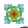
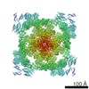
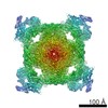
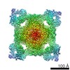
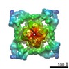
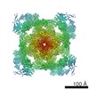
| Entry | Database: EMDB / ID: EMD-5014 | |||||||||
|---|---|---|---|---|---|---|---|---|---|---|
| Title | RyR1 in the closed state Ryanodine receptor 1 Ryanodine receptor 1 | |||||||||
 Map data Map data | 3D structure of RyR1 in the closed state. | |||||||||
 Sample Sample |
| |||||||||
 Keywords Keywords | ryanodine receptor / ion channel gating | |||||||||
| Biological species |  Oryctolagus cuniculus (rabbit) Oryctolagus cuniculus (rabbit) | |||||||||
| Method | single particle reconstruction / cryo EM / Resolution: 10.3 Å | |||||||||
 Authors Authors | Samso M / Wagenknecht T / Allen PD | |||||||||
 Citation Citation | Journal: Nat Struct Mol Biol / Year: 2005 Title: Internal structure and visualization of transmembrane domains of the RyR1 calcium release channel by cryo-EM. Authors: Montserrat Samsó / Terence Wagenknecht / P D Allen /  Abstract: RyR1 is an intracellular calcium channel with a central role in muscle contraction. We obtained a three-dimensional reconstruction of the RyR1 in the closed state at a nominal resolution of ...RyR1 is an intracellular calcium channel with a central role in muscle contraction. We obtained a three-dimensional reconstruction of the RyR1 in the closed state at a nominal resolution of approximately 10 A using cryo-EM. The cytoplasmic assembly consists of a series of interconnected tubular structures that merge into four columns that extend into the transmembrane assembly. The transmembrane assembly, which has at least six transmembrane alpha-helices per monomer, has four tilted rods that can be fitted with the inner helices of a closed K(+) channel atomic structure. The rods splay out at the lumenal side and converge into a dense ring at the cytoplasmic side. Another set of four rods emerges from this ring and shapes the inner part of the four columns. The resulting constricted axial structure provides direct continuity between cytoplasmic and transmembrane assemblies, and a possible mechanism for control of channel gating through conformational changes in the cytoplasmic assembly. | |||||||||
| History |
|
- Structure visualization
| Movie |
 Movie viewer Movie viewer |
|---|---|
| Structure viewer | EM map: SurfViewMolmilJmol/JSmol |
| Supplemental images |

- Downloads & links
-EMDB archive
| Map data | emd_5014.map.gz | 20.7 MB | EMDB map data format | |
|---|---|---|---|---|
| Header (meta data) | emd-5014-v30.xmlemd-5014.xml | 10.1 KB 10.1 KB | Display Display | EMDB header |
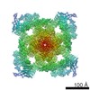
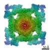
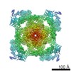
| Images | 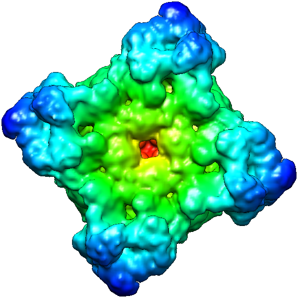 emd_5014_1.png emd_5014_1.png | 253.4 KB | ||
| Archive directory |  http://ftp.pdbj.org/pub/emdb/structures/EMD-5014ftp://ftp.pdbj.org/pub/emdb/structures/EMD-5014 http://ftp.pdbj.org/pub/emdb/structures/EMD-5014ftp://ftp.pdbj.org/pub/emdb/structures/EMD-5014 | HTTPS FTP |
-Related structure data
| Similar structure data |
|---|
-Links
| EMDB pages | EMDB (EBI/PDBe) / EMDataResource |
|---|
-Map
| File | Download / File: emd_5014.map.gz / Format: CCP4 / Size: 21.7 MB / Type: IMAGE STORED AS FLOATING POINT NUMBER (4 BYTES) | ||||||||||||||||||||||||||||||||||||||||||||||||||||||||||||||||||||
|---|---|---|---|---|---|---|---|---|---|---|---|---|---|---|---|---|---|---|---|---|---|---|---|---|---|---|---|---|---|---|---|---|---|---|---|---|---|---|---|---|---|---|---|---|---|---|---|---|---|---|---|---|---|---|---|---|---|---|---|---|---|---|---|---|---|---|---|---|---|
| Annotation | 3D structure of RyR1 in the closed state. | ||||||||||||||||||||||||||||||||||||||||||||||||||||||||||||||||||||
| Voxel size | X=Y=Z: 2.8 Å | ||||||||||||||||||||||||||||||||||||||||||||||||||||||||||||||||||||
| Density |
| ||||||||||||||||||||||||||||||||||||||||||||||||||||||||||||||||||||
| Symmetry | Space group: 1 | ||||||||||||||||||||||||||||||||||||||||||||||||||||||||||||||||||||
| Details | EMDB XML:
CCP4 map header:
| ||||||||||||||||||||||||||||||||||||||||||||||||||||||||||||||||||||
-Supplemental data
- Sample components
-Entire : ryanodine receptor 1
| Entire | Name: ryanodine receptor 1 |
|---|---|
| Components |
|
-Supramolecule #1000: ryanodine receptor 1
| Supramolecule | Name: ryanodine receptor 1 / type: sample / ID: 1000 / Oligomeric state: homotetramer / Number unique components: 1 |
|---|---|
| Molecular weight | Theoretical: 565 KDa |
-Supramolecule #1: ryanodine receptor isoform 1
| Supramolecule | Name: ryanodine receptor isoform 1 / type: organelle_or_cellular_component / ID: 1 / Name.synonym: RyR1 / Number of copies: 4 / Oligomeric state: Tetramer / Recombinant expression: No / Database: NCBI |
|---|---|
| Ref GO | divclassse qspanoncli ckpopupspa nclassgree n(this)spandata popltspanc lassquotlo adingbarqu otgtltimgs rcquotimgl oadinggifq uotdecodin gquotasync quotgtltsp angtdataur lajaxphp?m odetaxoamp ... divclassse qspanoncli ckpopupspa nclassgree n(this)spandata popltspanc lassquotlo adingbarqu otgtltimgs rcquotimgl oadinggifq uotdecodin gquotasync quotgtltsp angtdataur lajaxphp?m odetaxoamp kGO3A00052 19ampajax1 classpoptr giGO000521 9ispandiv |
| Ref INTERPRO | divclassse qspanoncli ckpopupspa nclassgree n(this)spandata popltspanc lassquotlo adingbarqu otgtltimgs rcquotimgl oadinggifq uotdecodin gquotasync quotgtltsp angtdataur lajaxphp?m odetaxoamp ... divclassse qspanoncli ckpopupspa nclassgree n(this)spandata popltspanc lassquotlo adingbarqu otgtltimgs rcquotimgl oadinggifq uotdecodin gquotasync quotgtltsp angtdataur lajaxphp?m odetaxoamp kIPR000699 ampajax1cl asspoptrgi IPR000699i spandiv |
| Source (natural) | Organism: Oryctolagus cuniculus (rabbit) / synonym: rabbit / Tissue: fast twitch skeletal muscle / Organelle: sarcoplasmic reticulum / Location in cell: sarcoplasmic reticulum membrane |
| Molecular weight | Experimental: 565 KDa / Theoretical: 565 KDa |
-Experimental details
-Structure determination
| Method | cryo EM |
|---|---|
 Processing Processing | single particle reconstruction |
| Aggregation state | particle |
-Sample preparation
| Concentration | 2.00 mg/mL |
|---|---|
| Buffer | pH: 7.4 Details: 20 mM Na-MOPS pH 7.4, 0.9 M NaCl, 0.5% (w/v) CHAPS, 2 mM DTT, 2 mM EGTA |
| Grid | Details: 300 mesh holey copper grids |
| Vitrification | Cryogen name: ETHANE / Instrument: HOMEMADE PLUNGER Details: Vitrification instrument: two-side blotting plunger Method: Blot for 2 seconds before plunging |
- Electron microscopy
| Microscope | FEI TECNAI F20 |
|---|---|
| Electron beam | Acceleration voltage: 200 kV / Electron source: FIELD EMISSION GUN |
| Electron optics | Illumination mode: FLOOD BEAM / Imaging mode: BRIGHT FIELDBright-field microscopy / Cs: 2.0 mm / Nominal defocus max: 4.0 µm / Nominal defocus min: 2.5 µm / Nominal magnification: 50000 |
| Sample stage | Specimen holder: Side entry liquid nitrogen-cooled cryo specimen holder Specimen holder model: GATAN LIQUID NITROGEN |
| Temperature | Average: 87 K |
| Alignment procedure | Legacy - Astigmatism: objective lens astigmatism was corrected at 150,000 times magnification |
| Image recording | Category: FILM / Film or detector model: KODAK SO-163 FILM / Digitization - Scanner: ZEISS SCAI / Digitization - Sampling interval: 7.0 µm / Number real images: 818 / Average electron dose: 10 e/Å2 / Details: after scanning pixels were averaged 2x2 / Bits/pixel: 8 |
| Experimental equipment |  Model: Tecnai F20 / Image courtesy: FEI Company |
-Image processing
| CTF correction | Details: Each particle |
|---|---|
| Final reconstruction | Algorithm: OTHER / Resolution.type: BY AUTHOR / Resolution: 10.3 Å / Resolution method: OTHER / Software - Name: FREALIGN and SPIDER / Number images used: 25722 |
-Atomic model buiding 1
| Initial model | PDB ID: |
|---|---|
| Details | The inner helices of KcsA (residues 72-119) were fitted by manual docking to the inner helices of RyR1 using program Chimera |
| Refinement | Space: REAL / Protocol: RIGID BODY FIT |

