 Movie
Movie Controller

[English] 日本語
 Yorodumi
- EMDB-4039: Maltose binding protein genetically fused to dodecameric glutamin... -
+ Open data

- Basic information
| Entry | Database: EMDB / ID: EMD-4039 | |||||||||
|---|---|---|---|---|---|---|---|---|---|---|
| Title | Maltose binding protein genetically fused to dodecameric glutamine synthetase | |||||||||
 Map data Map data | Maltose-binding protein fused to dodecameric glutamine synthetase | |||||||||
 Sample Sample |
| |||||||||
| Function / homology |  Function and homology information Function and homology information glutamine synthetase / glutamine biosynthetic process / glutamine synthetase activity / carbohydrate transmembrane transporter activity / outer membrane-bounded periplasmic space / ATP binding / metal ion binding / cytoplasm glutamine synthetase / glutamine biosynthetic process / glutamine synthetase activity / carbohydrate transmembrane transporter activity / outer membrane-bounded periplasmic space / ATP binding / metal ion binding / cytoplasmSimilarity search - Function | |||||||||
| Biological species |  Escherichia coli (E. coli) / Salmonella typhi (bacteria) / Escherichia coli O157:H7 (bacteria) Escherichia coli (E. coli) / Salmonella typhi (bacteria) / Escherichia coli O157:H7 (bacteria) | |||||||||
| Method | single particle reconstruction / cryo EM / Resolution: 6.2 Å | |||||||||
 Authors Authors | Coscia F / Petosa C / Schoehn G | |||||||||
| Funding support |  France, 1 items France, 1 items
| |||||||||
 Citation Citation | Journal: Sci Rep / Year: 2016 Title: Fusion to a homo-oligomeric scaffold allows cryo-EM analysis of a small protein. Authors: Francesca Coscia / Leandro F Estrozi / Fabienne Hans / Hélène Malet / Marjolaine Noirclerc-Savoye / Guy Schoehn / Carlo Petosa / Abstract: Recent technical advances have revolutionized the field of cryo-electron microscopy (cryo-EM). However, most monomeric proteins remain too small (<100 kDa) for cryo-EM analysis. To overcome this limitation, we explored a strategy whereby a monomeric target protein is genetically fused to a homo-oligomeric scaffold protein and the junction optimized to allow the target to adopt the scaffold symmetry, thereby generating a chimeric particle suitable for cryo-EM. To demonstrate the concept, we fused maltose-binding protein (MBP), a 40 kDa monomer, to glutamine synthetase, a dodecamer formed by two hexameric rings. Chimeric constructs with different junction lengths were screened by biophysical analysis and negative-stain EM. The optimal construct yielded a cryo-EM reconstruction that revealed the MBP structure at sub-nanometre resolution. These findings illustrate the feasibility of using homo-oligomeric scaffolds to enable cryo-EM analysis of monomeric proteins, paving the way for applying this strategy to challenging structures resistant to crystallographic and NMR analysis. | |||||||||
| History |
|
- Structure visualization
| Movie |
Movie viewer |
|---|---|
| Structure viewer | EM map: SurfViewMolmilJmol/JSmol |
| Supplemental images |

- Downloads & links
-EMDB archive
| Map data | emd_4039.map.gz | 191.9 MB | EMDB map data format | |
|---|---|---|---|---|
| Header (meta data) | emd-4039-v30.xmlemd-4039.xml | 15.5 KB 15.5 KB | Display Display | EMDB header |
| Images | 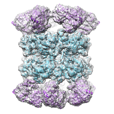 emd_4039.png emd_4039.png | 216 KB | ||
| Archive directory |  http://ftp.pdbj.org/pub/emdb/structures/EMD-4039ftp://ftp.pdbj.org/pub/emdb/structures/EMD-4039 http://ftp.pdbj.org/pub/emdb/structures/EMD-4039ftp://ftp.pdbj.org/pub/emdb/structures/EMD-4039 | HTTPS FTP |
-Related structure data
| Related structure data |  5ldfMC M: atomic model generated by this map C: citing same article ( |
|---|---|
| Similar structure data |

-Links
| EMDB pages | EMDB (EBI/PDBe) / EMDataResource |
|---|---|
| Related items in Molecule of the Month |


-Map
| File | Download / File: emd_4039.map.gz / Format: CCP4 / Size: 244.1 MB / Type: IMAGE STORED AS FLOATING POINT NUMBER (4 BYTES) | ||||||||||||||||||||||||||||||||||||||||||||||||||||||||||||
|---|---|---|---|---|---|---|---|---|---|---|---|---|---|---|---|---|---|---|---|---|---|---|---|---|---|---|---|---|---|---|---|---|---|---|---|---|---|---|---|---|---|---|---|---|---|---|---|---|---|---|---|---|---|---|---|---|---|---|---|---|---|
| Annotation | Maltose-binding protein fused to dodecameric glutamine synthetase | ||||||||||||||||||||||||||||||||||||||||||||||||||||||||||||
| Voxel size | X=Y=Z: 0.8155 Å | ||||||||||||||||||||||||||||||||||||||||||||||||||||||||||||
| Density |
| ||||||||||||||||||||||||||||||||||||||||||||||||||||||||||||
| Symmetry | Space group: 1 | ||||||||||||||||||||||||||||||||||||||||||||||||||||||||||||
| Details | EMDB XML:
CCP4 map header:
| ||||||||||||||||||||||||||||||||||||||||||||||||||||||||||||
-Supplemental data
- Sample components
-Entire : Maltose-binding protein genetically fused to glutamine synthetase
| Entire | Name: Maltose-binding protein genetically fused to glutamine synthetase |
|---|---|
| Components |
|
-Supramolecule #1: Maltose-binding protein genetically fused to glutamine synthetase
| Supramolecule | Name: Maltose-binding protein genetically fused to glutamine synthetase type: complex / ID: 1 / Parent: 0 / Macromolecule list: all |
|---|---|
| Source (natural) | Organism: Escherichia coli (E. coli) |
| Recombinant expression | Organism: Escherichia coli (E. coli) / Recombinant plasmid: pETM-11 |
| Molecular weight | Theoretical: 1.11 MDa |
-Macromolecule #1: Glutamine synthetase
| Macromolecule | Name: Glutamine synthetase / type: protein_or_peptide / ID: 1 / Number of copies: 12 / Enantiomer: LEVO / EC number: glutamine synthetase |
|---|---|
| Source (natural) | Organism: Salmonella typhi (bacteria) |
| Molecular weight | Theoretical: 51.586266 KDa |
| Recombinant expression | Organism: Escherichia coli (E. coli) |
| Sequence | String: EHVLTMLNEH EVKFVDLRFT DTKGKEQHVT IPAHQVNAEF FEEGKMFDGS SIGGWKGINE SDMVLMPDAS TAVIDPFFAD STLIIRCDI LEPGTLQGYD RDPRSIAKRA EDYLRATGIA DTVLFGPEPE FFLFDDIRFG ASISGSHVAI DDIEGAWNSS T KYEGGNKG ...String: EHVLTMLNEH EVKFVDLRFT DTKGKEQHVT IPAHQVNAEF FEEGKMFDGS SIGGWKGINE SDMVLMPDAS TAVIDPFFAD STLIIRCDI LEPGTLQGYD RDPRSIAKRA EDYLRATGIA DTVLFGPEPE FFLFDDIRFG ASISGSHVAI DDIEGAWNSS T KYEGGNKG HRPGVKGGYF PVPPVDSAQD IRSEMCLVME QMGLVVEAHH HEVATAGQNE VATRFNTMTK KADEIQIYKY VV HNVAHRF GKTATFMPKP MFGDNGSGMH CHMSLAKNGT NLFSGDKYAG LSEQALYYIG GVIKHAKAIN ALANPTTNSY KRL VPGYEA PVMLAYSARN RSASIRIPVV ASPKARRIEV RFPDPAANPY LCFAALLMAG LDGIKNKIHP GEPMDKNLYD LPPE EAKEI PQVAGSLEEA LNALDLDREF LKAGGVFTDE AIDAYIALRR EEDDRVRMTP HPVEFELYYS V |
-Macromolecule #2: Maltose-binding periplasmic protein
| Macromolecule | Name: Maltose-binding periplasmic protein / type: protein_or_peptide / ID: 2 / Number of copies: 12 / Enantiomer: LEVO |
|---|---|
| Source (natural) | Organism: Escherichia coli O157:H7 (bacteria) |
| Molecular weight | Theoretical: 40.753152 KDa |
| Recombinant expression | Organism: Escherichia coli (E. coli) |
| Sequence | String: KIEEGKLVIW INGDKGYNGL AEVGKKFEKD TGIKVTVEHP DKLEEKFPQV AATGDGPDII FWAHDRFGGY AQSGLLAEIT PDKAFQDKL YPFTWDAVRY NGKLIAYPIA VEALSLIYNK DLLPNPPKTW EEIPALDKEL KAKGKSALMF NLQEPYFTWP L IAADGGYA ...String: KIEEGKLVIW INGDKGYNGL AEVGKKFEKD TGIKVTVEHP DKLEEKFPQV AATGDGPDII FWAHDRFGGY AQSGLLAEIT PDKAFQDKL YPFTWDAVRY NGKLIAYPIA VEALSLIYNK DLLPNPPKTW EEIPALDKEL KAKGKSALMF NLQEPYFTWP L IAADGGYA FKYENGKYDI KDVGVDNAGA KAGLTFLVDL IKNKHMNADT DYSIAEAAFN KGETAMTING PWAWSNIDTS KV NYGVTVL PTFKGQPSKP FVGVLSAGIN AASPNKELAK EFLENYLLTD EGLEAVNKDK PLGAVALKSY EEELAKDPRI AAT MENAQK GEIMPNIPQM SAFWYAVRTA VINAASGRQT VDEALKDAQT RITK |
-Experimental details
-Structure determination
| Method | cryo EM |
|---|---|
 Processing Processing | single particle reconstruction |
| Aggregation state | particle |
-Sample preparation
| Concentration | 0.5 mg/mL | |||||||||||||||
|---|---|---|---|---|---|---|---|---|---|---|---|---|---|---|---|---|
| Buffer | pH: 8 Component:
| |||||||||||||||
| Grid | Model: Quantifoil / Material: COPPER/RHODIUM / Mesh: 400 / Support film - Material: CARBON / Support film - topology: HOLEY / Pretreatment - Type: GLOW DISCHARGE | |||||||||||||||
| Vitrification | Cryogen name: ETHANE / Chamber humidity: 100 % / Chamber temperature: 290 K / Instrument: FEI VITROBOT MARK IV |
- Electron microscopy
| Microscope | FEI TECNAI F30 |
|---|---|
| Electron beam | Acceleration voltage: 300 kV / Electron source: FIELD EMISSION GUN |
| Electron optics | Illumination mode: FLOOD BEAM / Imaging mode: BRIGHT FIELDBright-field microscopy / Nominal defocus max: 3.5 µm / Nominal defocus min: 1.0 µm |
| Sample stage | Cooling holder cryogen: NITROGEN |
| Details | Polara Top entry |
| Image recording | Film or detector model: GATAN K2 SUMMIT (4k x 4k) / Detector mode: SUPER-RESOLUTION / Number real images: 165 / Average exposure time: 6.0 sec. / Average electron dose: 25.0 e/Å2 |
| Experimental equipment |  Model: Tecnai F30 / Image courtesy: FEI Company |
-Image processing
| Particle selection | Number selected: 39167 |
|---|---|
| CTF correction | Software: (Name: CTFFIND (ver. 3), Bsoft) |
| Startup model | Type of model: OTHER Details: Reference models were generated by angular reconstitution using IMAGIC and volumes refined by projection matching using SPIDER. As a control to check for possible model bias, we generated an ...Details: Reference models were generated by angular reconstitution using IMAGIC and volumes refined by projection matching using SPIDER. As a control to check for possible model bias, we generated an alternative ab initio reference model using RIco, which uses symmetry adapted functions. |
| Initial angle assignment | Type: ANGULAR RECONSTITUTION / Software - Name: IMAGIC |
| Final 3D classification | Software - Name: RELION |
| Final angle assignment | Type: PROJECTION MATCHING |
| Final reconstruction | Applied symmetry - Point group: D6 (2x6 fold dihedral) / Resolution.type: BY AUTHOR / Resolution: 6.2 Å / Resolution method: FSC 0.143 CUT-OFF / Software - Name: RELION / Number images used: 13847 |
-Atomic model buiding 1
| Refinement | Space: REAL / Protocol: RIGID BODY FIT |
|---|---|
| Output model | PDB-5ldf: |
